SPLEEN
Structure
The spleen has a slightly oval shape. It is covered by a weak capsule that protects the organ whilst allowing it to expand in size.
The outer surface of the spleen can be anatomically divided into two:
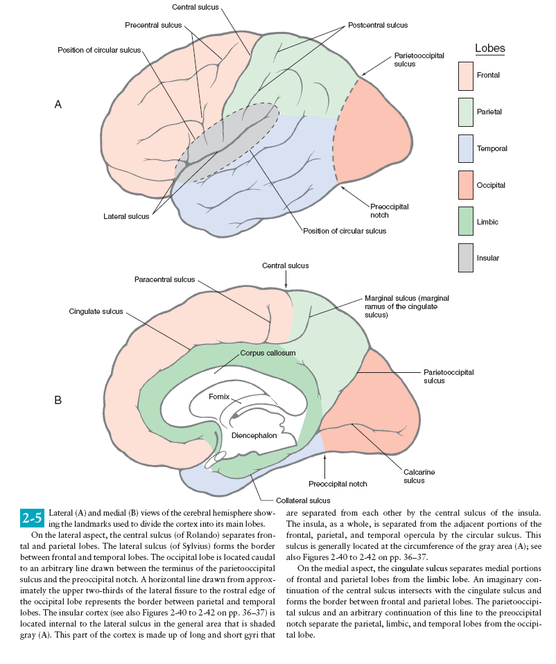
Saggital Section of Brainstem
The brain stem consists of the medulla oblongata, pons and midbrain. It is
sited in the posterior cranial fossa, and its ventral surface lies on the clivus.
It contains numerous intrinsic neurone cell bodies and their processes, some
of which are the brain stem homologues of spinal neuronal groups. These
include the sites of termination and cells of origin of axons that enter or leave
the brain stem through the cranial nerves. They provide the sensory, motor
and autonomic innervation of structures that are mostly in the head and
SPLEEN
The spleen is an organ located in the upper left abdomen, and is roughly the size of a clenched fist. In the adult, the spleen functions mainly as a blood filter, removing old red blood cells. It also plays a role in both cell-mediated and humoral immune responses.
In this article, we shall look at the anatomy of the spleen – its anatomical position, structure and vasculature.
By TeachMeSeries Ltd (2023)
MUSCLES OF THIGH WITH GLUTEUS MAXIMUS
The gluteal region is an anatomical area located posteriorly to the pelvic girdle, at the proximal end of the femur. The muscles in this region move the lower limb at the hip joint.
The muscles of the gluteal region can be broadly divided into two groups:
Saggital Section of Brain
https://youtu.be/2ztESvDnxT0
24. kidney
The kidneys are two reddish-brown bean-shaped organs found in vertebrates.
ARTERY & VEIN
What are blood vessels?
Blood vessels are channels that carry blood throughout your body. They form a closed loop, like a circuit, that begins and ends at your heart. Together, the heart vessels and blood vessels form your circulatory system. Your body contains about 60,000 miles of blood vessels.
There are three types of blood vessels:
KIDNEY WITH URETER
The kidneys are bilateral organs placed retroperitoneally in the upper left and right abdominal quadrants and are part of the urinary system. Their shape resembles a bean, where we can describe the superior and inferior poles, as well as the major convexity pointed laterally, and the minor concavity pointed medially.
The main function of the kidney is to eliminate excess bodily fluid, salts and byproducts of metabolism – this makes kidneys key in the regulation of acid-base balance, blood pressure, and many other homeostatic parameters.
Key facts about the kidney
TESTIES
The testes and epididymis are paired structures, located within the scrotum. The testes are the site of sperm production and hormone synthesis, while the epididymis has a role in the storage of sperm.
In this article, we shall look at the anatomy of the testes and epididymis – their structure, vasculature, innervation and clinical correlations.