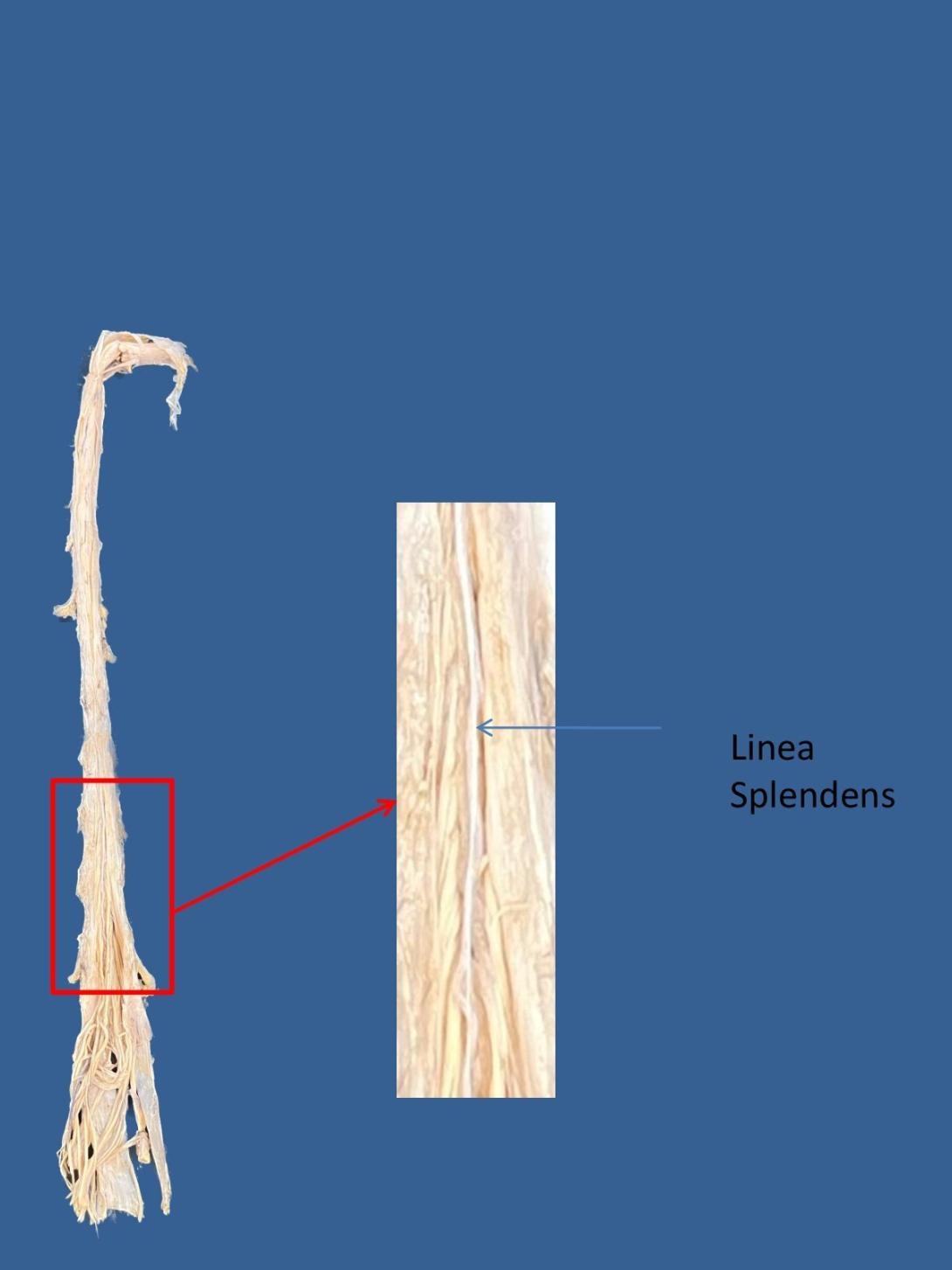
The spinal cord is a tubular bundle of nervous tissue and supporting cells that extends from the brainstem to the lumbar vertebrae. Together, the spinal cord and the brain form the central nervous system.
In this article, we shall examine the macroscopic anatomy of the spinal cord – its structure, membranous coverings and blood supply.
For information regarding the internal structure of the spinal cord, see the grey matter of the spinal cord.
Anatomical Position and Structure
The spinal cord is a cylindrical structure, greyish-white in colour. It has a relatively simple anatomical course:
- The spinal cord arises cranially as a continuation of the medulla oblongata (part of the brainstem).
- It then travels inferiorly within the vertebral canal, surrounded by the spinal meninges containing cerebrospinal fluid.
- At the L2 vertebral level the spinal cord tapers off, forming the conus medullaris.
As a result of the termination of the spinal cord at L2, it occupies around two thirds of the vertebral canal. The spinal nerves that arise from the end of the spinal cord are bundled together, forming a structure known as the cauda equina.
During the course of the spinal cord, there are two points of enlargement. The cervical enlargement is located proximally, at the C4-T1 level. It represents the origin of the brachial plexus. Between T11 and L1 is the lumbar enlargement, representing the origin of the lumbar and sacral plexi.
The spinal cord is marked by two depressions on its surface. The anterior median fissure is a deep groove extending the length of the anterior surface of the spinal cord. On the posterior aspect there is a slightly shallower depression – the posterior median sulcus.
By TeachMeSeries Ltd (2023)

