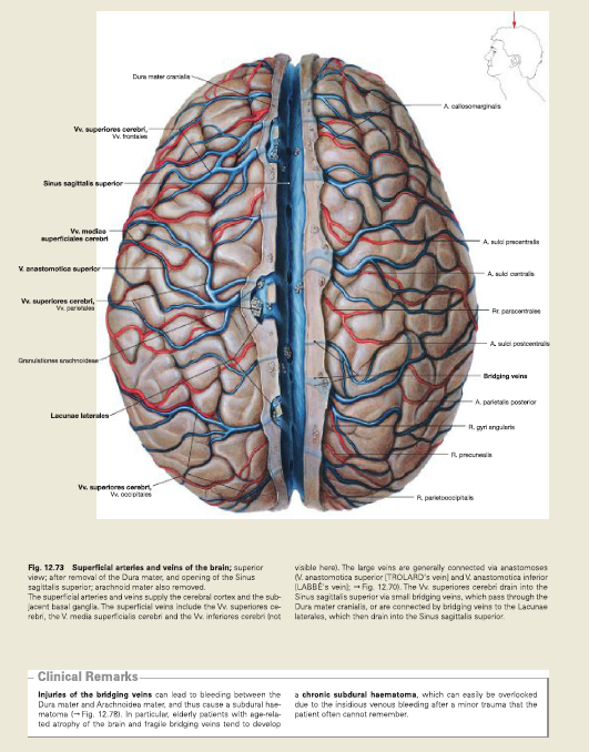
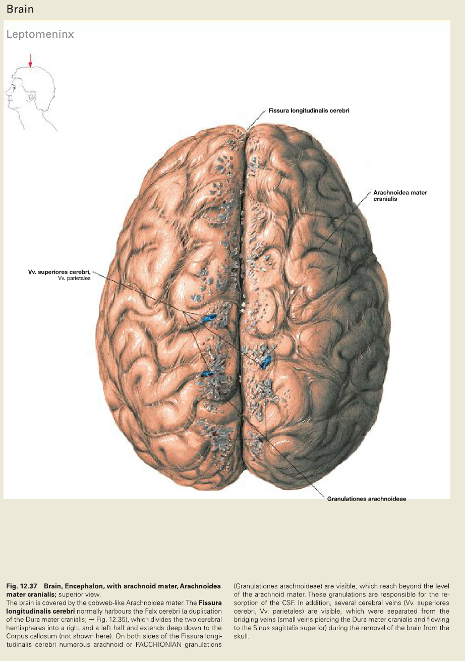
SKULL CAP
Skull cap bone
The calvarium, also known as the roof or skull cap, consists of three bones: Frontal bones. Parietal bones. Occipital bones.
SAGITTAL SECTION OF HUMERUS BONE
Bones are composed of bone matrix, which has both organic and inorganic components. Bone matrix is laid down by osteoblasts as collagen, also known as osteoid. Osteoid is hardened with inorganic salts, such as calcium and phosphate, and by the chemicals released from the osteoblasts through a process known as mineralization.
The basic microscopic unit of bone is an osteon (or Haversian system). Osteons are roughly cylindrical structures that can measure several millimeters long and around 0.2 mm in diameter.
SAGITTAL SECTION OF TIBIA BONE
The tibia is a medial and large long bone of the lower extremity, connecting the knee and ankle joints. It is considered to be the second largest bone in the body and it plays an important role in weight bearing.[1] Osteologic features of the tibia include medial and lateral condyles, the tibial plateau, the tibial tuberosity, the soleal line, the medial malleolus, and the fibular notch.
Osteologic Features
LIVER
Clinical Relevance: Percutaneous Liver Biopsy
A percutaneous liver biopsy is procedure used to obtain a sample of liver tissue. A needle is inserted through the skin to access the liver.
The biopsy is required in several clinical scenarios:
SUPERIOR THORACIC INLET
The superior thoracic aperture, also known as the thoracic outlet, or thoracic inlet refers to the opening at the top of the thoracic cavity. It is also clinically referred to as the thoracic outlet, in the case of thoracic outlet syndrome
Apart from the diaphragm, the list of structures that pass through the inferior thoracic outlet is best described by considering the various diaphragmatic apertures: aortic hiatus (T12) (not a true aperture) esophageal hiatus (T10) vena caval foramen (T8)

