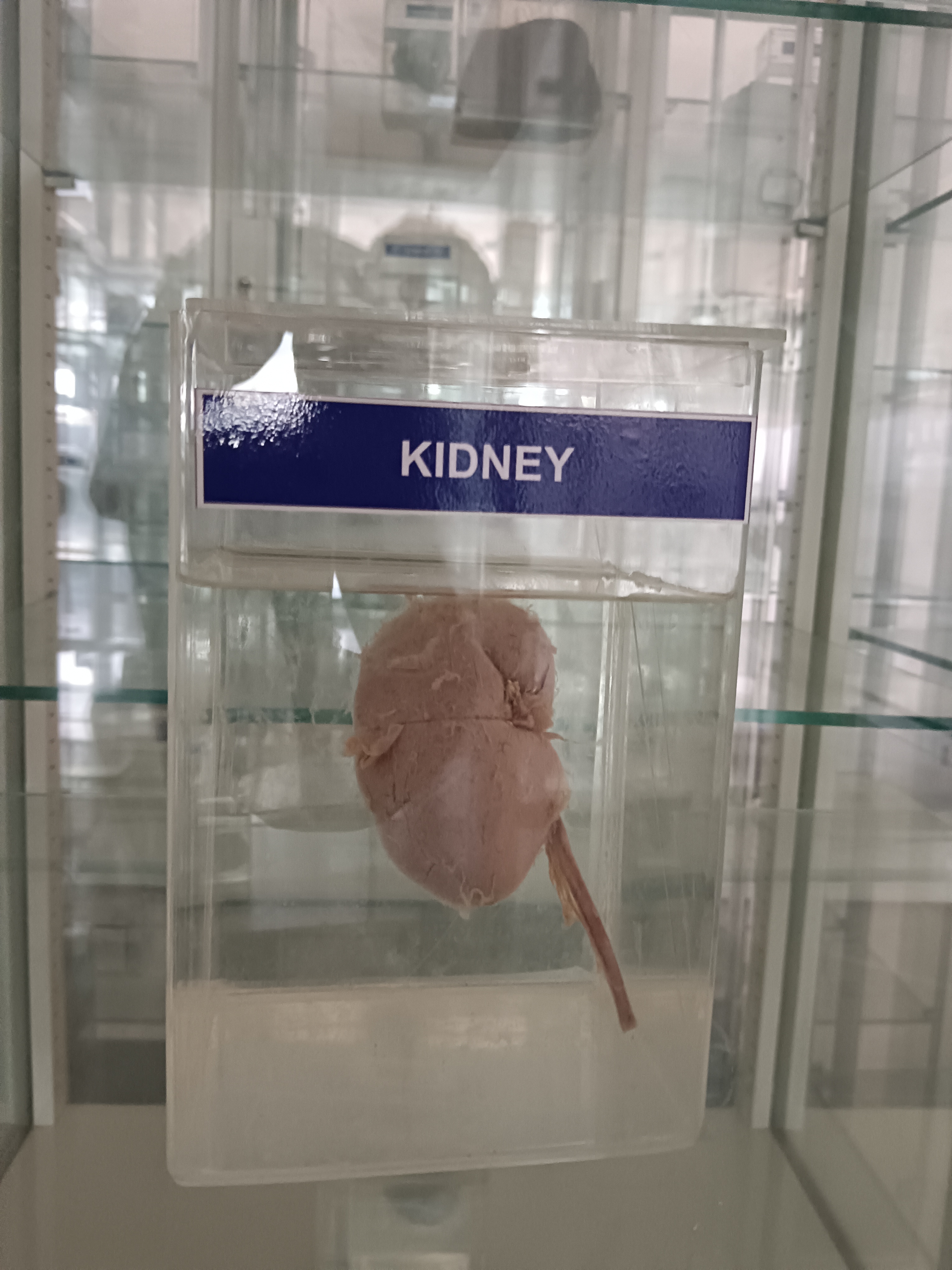
The kidneys are a pair of bean-shaped organs located in the abdomen, just below the ribcage, and on either side of the spine. The kidneys are an important part of the urinary system and play a vital role in filtering waste products and excess water from the blood to produce urine.
- Renal capsule: A thin layer of connective tissue that covers the outer surface of the kidney.
- Cortex: The outer layer of the kidney that contains the glomeruli, proximal and distal tubules, and the renal corpuscles.
- Medulla: The inner layer of the kidney that contains the loops of Henle and the collecting ducts.
- Renal papilla: The tip of the renal pyramid, where urine exits the kidney.
- Renal pelvis: A funnel-shaped structure that collects urine from the kidney and transports it to the ureter.
- Calyx: A cup-like structure that collects urine from the renal papilla and transports it to the renal pelvis.
- Nephron: The functional unit of the kidney, which consists of the glomerulus, proximal and distal tubules, and the loop of Henle.
- Renal artery and vein: Blood vessels that supply and drain the kidney.
- Ureter: A tube that carries urine from the kidney to the bladder.
The kidneys are innervated by both the sympathetic and parasympathetic nervous systems.
The sympathetic nerves originate from the thoracic and lumbar spinal cord segments and travel through the sympathetic ganglia to reach the kidneys. These nerves help regulate the blood flow to the kidneys and control the release of renin, a hormone that helps regulate blood pressure.
The parasympathetic nerves, which originate from the vagus nerve, also have a role in kidney function. These nerves help regulate the blood flow to the kidneys and control the release of urine.
In addition to the autonomic nervous system, the kidneys are also innervated by sensory nerves that transmit pain and other sensations from the kidney to the central nervous system.
Overall, the nervous system plays an important role in regulating kidney function, blood flow, and blood pressure. Dysfunction in the nervous system can contribute to various kidney disorders, including hypertension and kidney disease.