Specimen Image
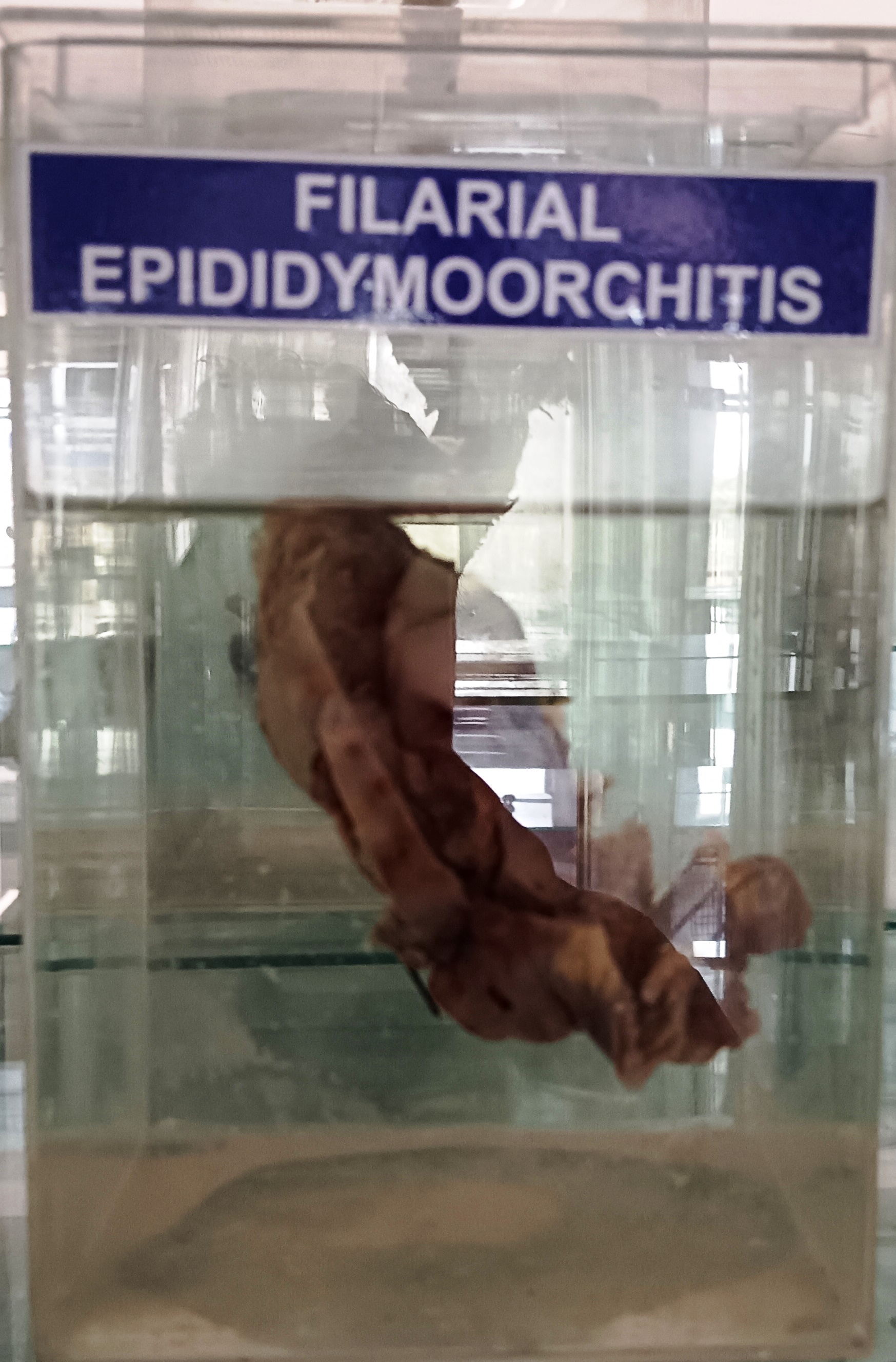
Filarial epididymoorchitis is a condition characterized by inflammation of the epididymis and testicles due to infection with filarial parasites. Filarial parasites are tiny, thread-like worms that are transmitted to humans through the bite of infected mosquitoes.
The symptoms of filarial epididymoorchitis can vary but commonly include swelling, pain, and tenderness in the affected testicle, as well as fever and chills. In some cases, the lymph nodes in the groin may also become swollen and tender.
The treatment of filarial epididymoorchitis typically involves the use of medication to kill the filarial parasites and reduce inflammation. The medications used may include:
- Albendazole: It is an antiparasitic medication that works by killing the filarial parasites in the body.
- Ivermectin: It is another antiparasitic medication that can be used in combination with albendazole to kill the filarial parasites.
- Doxycycline: It is an antibiotic medication that is often used in combination with antiparasitic drugs to kill the filarial parasites and reduce inflammation.
Rack Number
Specimen Number
41