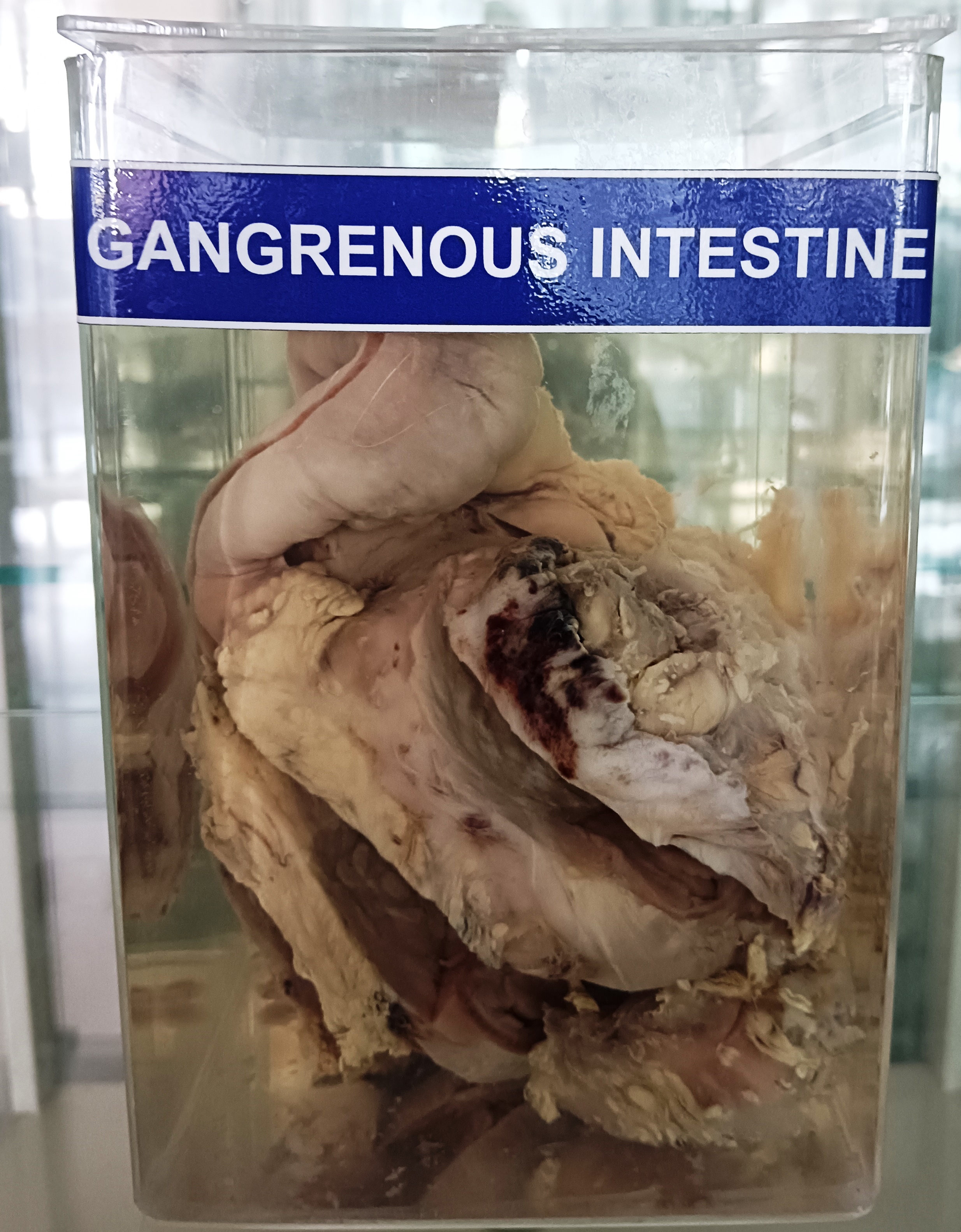
Gangrene of the intestine is a serious medical condition that arises when the blood supply to a portion of the intestine is compromised, leading to tissue death and decay. The condition can arise due to a variety of factors, including blockages in the blood vessels, infections, and trauma.
The intestine is a long, muscular tube that runs from the stomach to the anus. It is divided into two major sections: the small intestine and the large intestine. The small intestine is responsible for the majority of nutrient absorption, while the large intestine absorbs water and electrolytes from the undigested food material.
Pathologically, gangrene of the intestine is characterized by the death and decay of the intestinal tissues, which can lead to the release of toxins and bacteria into the bloodstream. This can cause a range of symptoms, including severe abdominal pain, fever, nausea, vomiting, and diarrhea. In severe cases, gangrene of the intestine can lead to sepsis, a life-threatening condition that can cause organ failure and death.
Treatment for gangrene of the intestine typically involves surgery to remove the affected portion of the intestine, along with the administration of antibiotics to control infection. In some cases, the remaining healthy intestine may be reconnected, while in other cases, a temporary or permanent colostomy or ileostomy may be required to allow for the elimination of waste.
Prevention of gangrene of the intestine involves managing risk factors such as diabetes, smoking, and obesity, and seeking prompt medical attention for symptoms such as abdominal pain, bloating, and diarrhea.