Pyelonephritis is an infection of the kidney, which is caused by bacteria that enter the kidney through the ureters, the tubes that carry urine from the bladder to the kidneys. The infection can be either acute or chronic and can affect one or both kidneys.
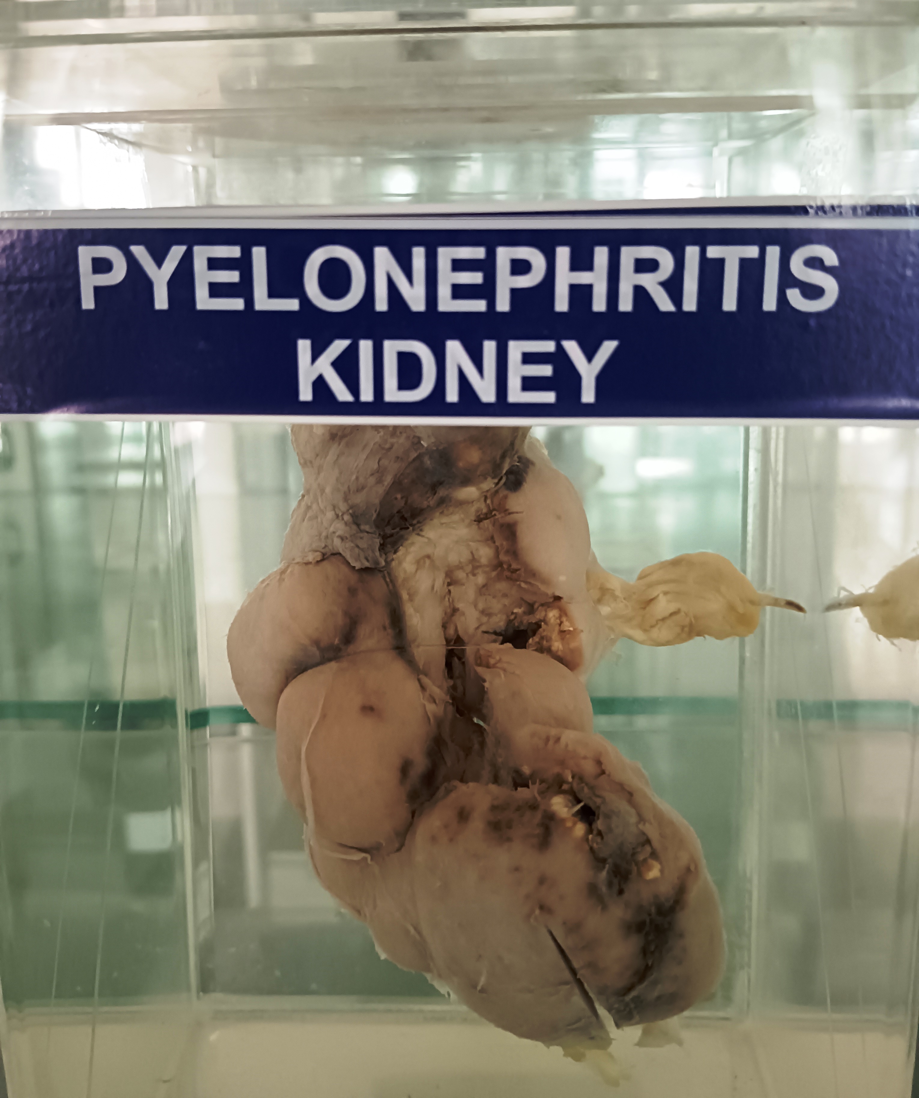
Pathologically, pyelonephritis is characterized by inflammation and swelling of the kidney tissue. In acute pyelonephritis, the kidney may appear enlarged and congested, with areas of suppuration or pus formation. The renal pelvis and calyces may be dilated, and there may be pus and debris in the urine.
In chronic pyelonephritis, the kidney may appear smaller and scarred, with atrophy of the renal parenchyma. The renal pelvis and calyces may be dilated and filled with pus, and there may be fibrosis and scarring of the kidney tissue.
The pathogenesis of pyelonephritis involves the ascent of bacteria from the lower urinary tract into the upper urinary tract, where they can infect the kidneys. Factors that increase the risk of pyelonephritis include urinary tract obstructions, vesicoureteral reflux, neurogenic bladder, and pregnancy.
Anatomy-wise, the kidney is a bean-shaped organ located in the retroperitoneal space, on either side of the vertebral column. It receives blood supply from the renal artery and drains into the renal vein. The urine produced by the kidney is transported to the bladder via the ureters.