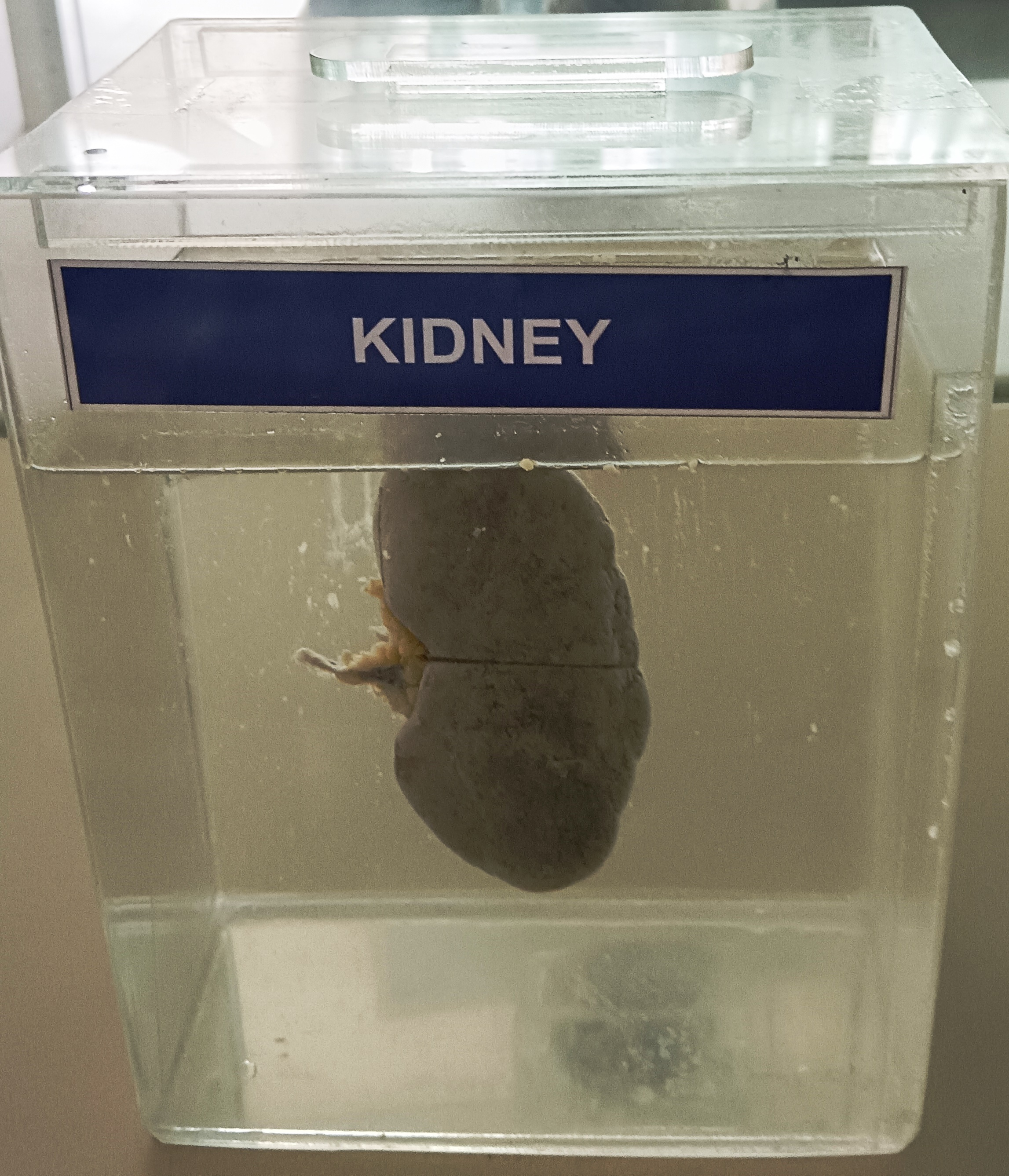
The kidneys are two bean-shaped organs located in the retroperitoneal space, on either side of the vertebral column. They are situated posteriorly to the peritoneum, behind the abdominal cavity, and are positioned higher on the left side than the right due to the liver's presence on the right side.
Each kidney is about 10-12 cm in length and weighs around 120-150 grams in adults. The kidney has a convex lateral border and a concave medial border, which forms the renal hilum, where the renal vessels, ureter, and nerves enter and exit the kidney. The renal hilum is located approximately at the level of the 1st or 2nd lumbar vertebra.
The kidney is made up of two main regions: the outer renal cortex and the inner renal medulla. The renal cortex is the outer layer of the kidney and contains the glomeruli (the filtering units of the kidney), the proximal and distal convoluted tubules, and the cortical collecting ducts. The renal medulla is the inner region of the kidney and contains the renal pyramids, which are conical-shaped structures that contain the loops of Henle and collecting ducts. The renal medulla is divided into several renal lobes, each containing a renal pyramid and the overlying cortex.
The renal pelvis is a funnel-shaped structure that collects urine from the collecting ducts and renal calyces, which are cup-shaped structures that collect urine from the renal papillae. The renal pelvis then narrows to form the ureter, which carries urine from the kidney to the bladder.
The blood supply to the kidney is via the renal artery, which branches into segmental arteries, which then divide into interlobar arteries. The interlobar arteries run along the boundary between the renal cortex and medulla and give rise to the arcuate arteries, which form the interlobular arteries that supply the glomeruli and cortical portions of the kidney.