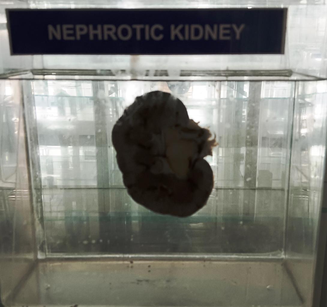
Nephrotic syndrome is a condition characterized by the presence of excess protein in the urine due to damage to the glomeruli, the tiny filtering units in the kidney. Here are some of the important anatomical points related to a nephrotic kidney:
https://www.youtube.com/watch?v=2tQDAjo5Utc
- Glomeruli: The glomeruli are tiny blood vessels in the kidney that filter waste products and excess fluid from the blood. In nephrotic syndrome, the glomeruli become damaged, allowing large amounts of protein to leak into the urine.
- Renal cortex: The outer layer of the kidney is called the renal cortex. It contains the glomeruli and the proximal and distal tubules of the nephrons. In nephrotic syndrome, the renal cortex may appear swollen due to the accumulation of excess fluid.
- Renal medulla: The inner layer of the kidney is called the renal medulla. It contains the loops of Henle, the collecting ducts, and the renal pyramids. In nephrotic syndrome, the renal medulla may appear normal.
- Renal capsule: The kidney is surrounded by a fibrous capsule that provides protection and support. In nephrotic syndrome, the renal capsule may be stretched due to the accumulation of excess fluid.
- Renal pelvis: The renal pelvis is a funnel-shaped area at the center of the kidney where urine collects before it enters the ureter. In nephrotic syndrome, the renal pelvis may appear dilated due to the accumulation of excess fluid.
- Ureter: The ureter is a long, muscular tube that carries urine from the kidney to the bladder. In nephrotic syndrome, the ureter may appear normal.
- Bladder: The bladder is a muscular sac that stores urine until it is eliminated from the body. In nephrotic syndrome, the bladder may appear normal.
- Urethra: The urethra is a tube that carries urine from the bladder to the outside of the body. In nephrotic syndrome, the urethra may appear normal.
Rack Number
Specimen Number
7

