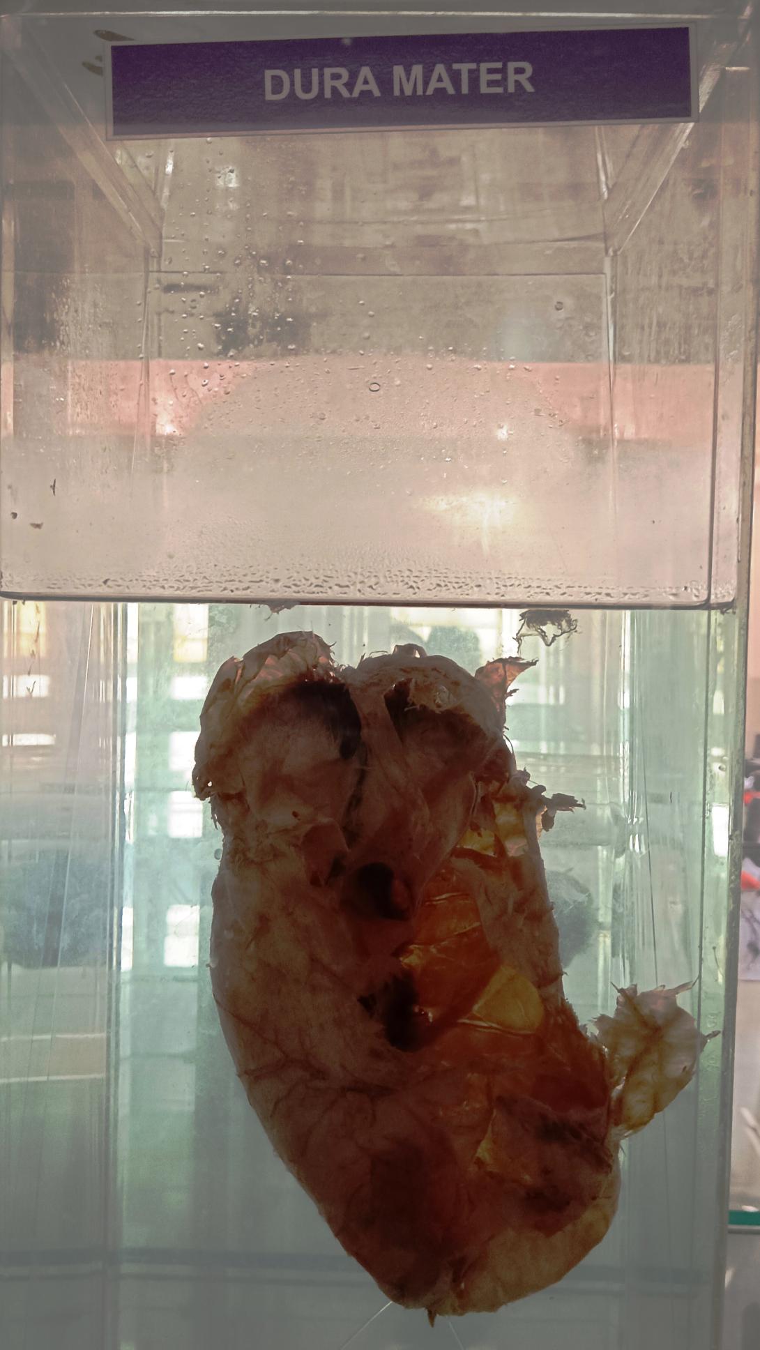
The dura mater is the outermost and toughest layer of the meninges, which are the membranes that cover and protect the brain and spinal cord. Here are some of the important anatomical points related to the dura mater:
- Structure: The dura mater is composed of two layers, the outer periosteal layer and the inner meningeal layer. These layers are fused together except in certain areas where they separate to form dural venous sinuses.
- Location: The dura mater is located just beneath the bones of the skull and vertebral column, forming a protective covering for the brain and spinal cord.
- Falx cerebri: The falx cerebri is a fold of the dura mater that separates the two cerebral hemispheres of the brain.
- Tentorium cerebelli: The tentorium cerebelli is a fold of the dura mater that separates the cerebellum from the occipital lobes of the brain.
- Falx cerebelli: The falx cerebelli is a small fold of the dura mater that separates the two hemispheres of the cerebellum.
- Dural venous sinuses: The dural venous sinuses are spaces between the two layers of the dura mater that drain blood and cerebrospinal fluid from the brain and spinal cord. Examples of dural venous sinuses include the superior sagittal sinus, transverse sinuses, and sigmoid sinuses.
- Foramen magnum: The foramen magnum is the large opening in the base of the skull through which the spinal cord enters and exits the skull. The dura mater forms a tough protective sheath around the spinal cord as it passes through the foramen magnum.
- Blood supply: The dura mater is supplied with blood by the meningeal branches of the external carotid artery and the vertebral artery.
- Innervation: The dura mater is innervated by the trigeminal nerve, the vagus nerve, and the first three cervical nerves. These nerves are responsible for providing sensation to the head and neck.
Rack Number
Specimen Number
1

