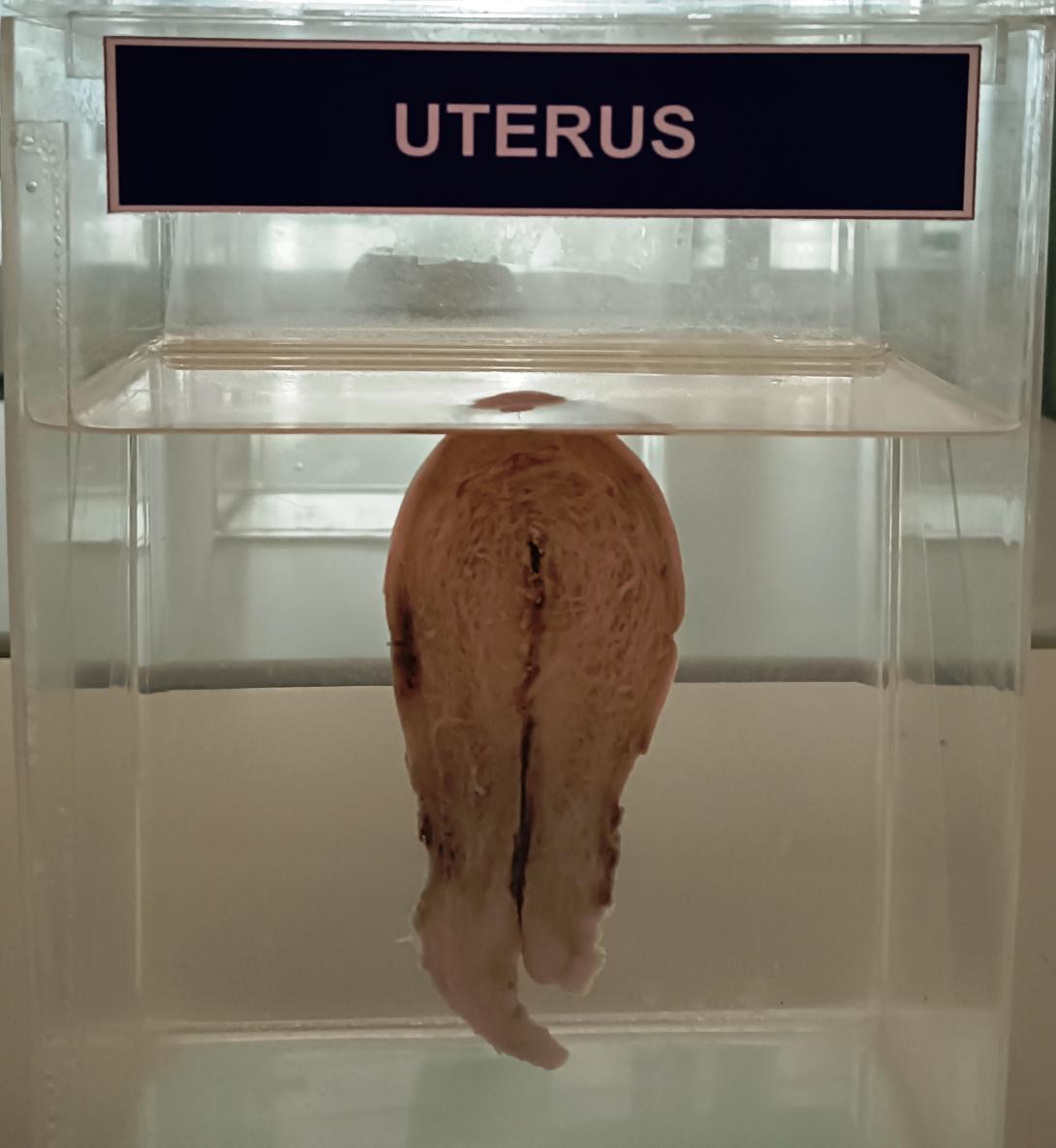
Support[edit]
Uterus covered by the broad ligament
The uterus is primarily supported by the pelvic diaphragm, perineal body, and urogenital diaphragm. Secondarily, it is supported by ligaments, including the peritoneal ligament and the broad ligament of uterus.[15]
Major ligaments[edit]
The uterus is held in place by several peritoneal ligaments, of which the following are the most important (there are two of each):
Name
From
To
Posterior cervix
Anterior face of sacrum
Side of the cervix
Side of the cervix
Axis[edit]
Normally, the human uterus lies in anteversion and anteflexion. In most women, the long axis of the uterus is bent forward on the long axis of the vagina, against the urinary bladder. This position is referred to as anteversion of the uterus. Furthermore, the long axis of the body of the uterus is bent forward at the level of the internal os with the long axis of the cervix. This position is termed anteflexion of the uterus.[16] The uterus assumes an anteverted position in 50% of women, a retroverted position in 25% of women, and a midposed position in the remaining 25% of women.[1]
Position[edit]
Uterus shown in position in the body
The uterus is located in the middle of the pelvic cavity, in the frontal plane (due to the broad ligament of the uterus). The fundus does not extend above the linea terminalis, while the vaginal part of the cervix does not extend below the interspinal line. The uterus is mobile and moves posteriorly under the pressure of a full bladder, or anteriorly under the pressure of a full rectum. If both are full, it moves upwards. Increased intra-abdominal pressure pushes it downwards. The mobility is conferred to it by a musculo-fibrous apparatus that consists of suspensory and sustentacular parts. Under normal circumstances, the suspensory part keeps the uterus in anteflexion and anteversion (in 90% of women) and keeps it "floating" in the pelvis. The meanings of these terms are described below:
1. Anteversion with slight anteflexion
2. Anteversion with marked anteflexion
3. Anteversion with retrocession
4. Retroversion
5. Retroversion with retroflexion
Distinction
More common
Less common
Position tipped
"Anteverted": Tipped forward
"Retroverted": Tipped backwards
Position of fundus
"Anteflexed": Fundus is pointing forward relative to the cervix
"Retroflexed": Fundus is pointing backward
The sustentacular part supports the pelvic organs and comprises the larger pelvic diaphragm in the back and the smaller urogenital diaphragm in the front.
The pathological changes of the position of the uterus are:
- retroversion/retroflexion, if it is fixed
- hyperanteflexion – tipped too forward; most commonly congenital, but may be caused by tumors
- anteposition, retroposition, lateroposition – the whole uterus is moved; caused by parametritis or tumors
- elevation, descensus, prolapse
- rotation (the whole uterus rotates around its longitudinal axis), torsion (only the body of the uterus rotates around)
- inversion
In cases where the uterus is "tipped", also known as retroverted uterus, the woman may have symptoms of pain during sexual intercourse, pelvic pain during menstruation, minor incontinence, urinary tract infections, fertility difficulties,[17] and difficulty using tampons. A pelvic examination by a doctor can determine if a uterus is tipped.[18]
Blood, lymph, and nerve supply supply[edit]
Diagram of uterine blood supply
The human uterus is supplied by arterial blood both from the uterine artery and the ovarian artery. Another anastomotic branch may also supply the uterus from anastomosis of these two arteries.
Afferent nerves supplying the uterus are T11 and T12. Sympathetic supply is from the hypogastric plexus and the ovarian plexus. Parasympathetic supply is from the S2, S3 and S4 nerves.

