
- Arnold-Chiari syndrome with hydrocephalus (Q07.-)
- congenital hydrocephalus (Q03.-)
- spina bifida with hydrocephalus (Q05.-)
Includes
- acquired hydrocephalus
The following code(s) above G91 contain annotation back-referencesthat may be applicable to G91:
- G00-G99Diseases of the nervous system
Clinical Information
- (hye-dro-sef-uh-lus) the abnormal buildup of cerebrospinal fluid in the ventricles of the brain.
- A disorder characterized by an abnormal increase of cerebrospinal fluid in the ventricles of the brain.
- Excessive accumulation of cerebrospinal fluid within the cranium which may be a congenital or acquired disorder; hydrocephalus ex-vacuo refers to ventricular dilation that occurs as a result of brain substance loss from cerebral infarction and other conditions.
- Excessive accumulation of cerebrospinal fluid within the cranium which may be associated with dilation of cerebral ventricles, intracranial hypertension; headache; lethargy; urinary incontinence; and ataxia.
- Excessive gathering of cerebrospinal fluid within the head bone
Hydrocephalus is the buildup of too much cerebrospinal fluid in the brain. Normally, this fluid cushions your brain. When you have too much, though, it puts harmful pressure on your brain.there are two kinds of hydrocephalus. Congenital hydrocephalus is present at birth. Causes include genetic problems and problems with how the fetus develops. An unusually large head is the main sign of congenital hydrocephalus. Acquired hydrocephalus can occur at any age. Causes can include head injuries, strokes, infections, tumors and bleeding in the brain. Symptoms of acquired hydrocephalus can include
- headache
- vomiting and nausea
- blurry vision
- balance problems
- bladder control problems
- thinking and memory problems
hydrocephalus can permanently damage the brain, causing problems with physical and mental development. If untreated, it is usually fatal. With treatment, many people lead normal lives with few limitations. Treatment usually involves surgery to insert a shunt. Medicine and rehabilitation therapy can also help.
- Hydrocephalus that results from head trauma, brain tumors, intracranial hemorrhage, or meningitis.
- The abnormal buildup of cerebrospinal fluid in the ventricles of the brain.
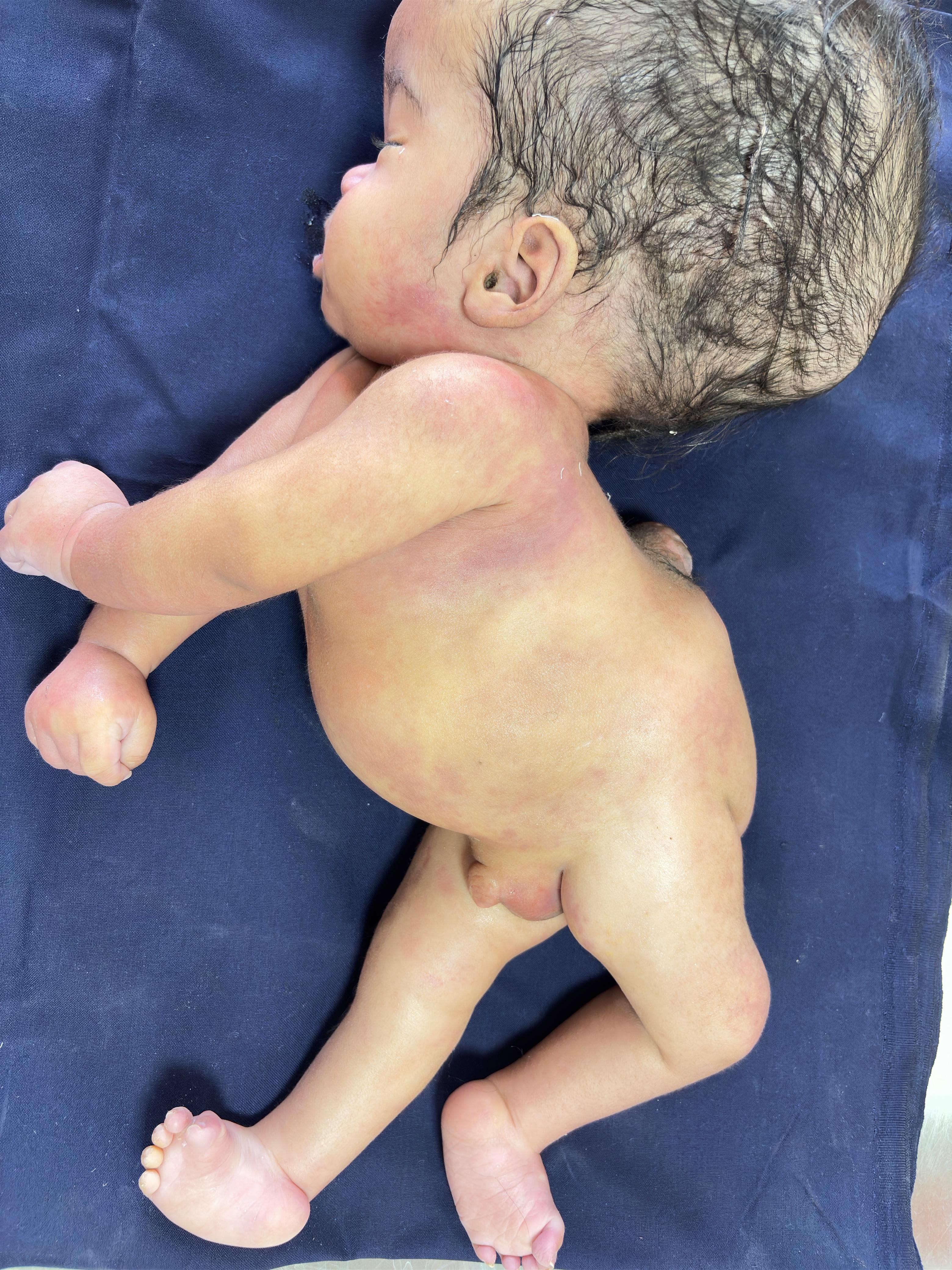
CLUB FOOT
- The club foot is an idiopathic or congenital deformity which occur in children, and its causes are yet unknown1 • 2. In case of the idiopathic club foot, the deformity is limited to the foot only while the rest of the musculoskeletal structure appears normal3• On the other hand the non-idiopathic clubfoot may be associated with other neuromuscular conditions like muscular dystrophy, diastrophic dwarfism, arthrogryposis and myelomeningocele3• However great effort is being made to trace an association between its genetic behavior and to link with other medical conditions, such as spina bifida, hydrocephalus and meningomyloceal etc
- The affected child acquires the combinations of the following deformities of the foot :
- ❖ Forefoot: The forefoot is adducted and supinated. 2 · 3
- ❖ Midfoot: High medial arched( caV\ls deformity).The first metatarsal is more plantar flexed than the fifth metatarsal which results in high arch appearance of the foot. 12, 3
- ❖ Hind foot: The hind foot is held in planter flexion and inversion (varus) position and the foot is twisted in towards the other foot. The heel is drawn up as in equinus deformity)2, 3, 15
MANAGEMENT:
There are different treatment approaches for club foot deformity11 . Conservative ( serial casting) method is the treatment of choice, other procedures are surgical and external fixators7 • 11 • Idiopathic clubfoot deformity is best treated with Conservative Techniques4 • 11 . In conservative treatments, the Ponsti method of treatment comes on the top of the list. While the French Physiotherapy comes next and is found to be equally effective4• 20• According to a long-term followup study, physical therapy without anesthesia or plaster casts was used to treat 338 cases of clubfoot (CF). The , technique was based on progressive sequential manipulations at birth. Varus deformity was reduced first and the equinus component of the club foot was manipulated later, followed up by gentle stretches, active physiotherapy and then a foot orthosis to secure its extent of rearrangement 11- 13 .
This technique accomplished 77% good and fair results. In challenging cases, subsequent surgical procedures were employed which showed 96% good and fair results 9 . Normal Clubfoot in baby PONSETI METHOD (Serial Casting): Ponseti method of serial casting is commonly executed by Orthopedic surgeons and Physical therapists. Gentle manipulation and serial casting is performed to rectify the foot position. This non-operative technique is put into practiced in 27 countries and has rapidly become the standard method of care as an initial treatment for clubfoot throughout the world, because it is efficient, secure and economical6 • 7 • 11 • 19•
The basic idea of this technique was developed by Dr. Ignacio Ponseti in the 1940s in which a weekly manipulation of the foot allows collagen relaxation of contracted ligaments, capsules and tendons, which .results in remodeling of the joint surfaces 11
• 12. Thus in this procedure the need for surgical correction, in many cases, can be J. Baqai Med. Univ. deformity, the normal position is maintained by particular footwear i.e, Ponseti FAO (Foot Abduction Orthosis) and regular phys'ical therapy exercise (stretching)12. This conservative method results good when treated within the age of 3-6 months4 - 12.
After the age of 5-6 months the ligaments get stiffer and child may require surgical correction 12. Method and Sequence of Manipulation of the CTEV: • Ponseti method of serial casting is recommended within 2-3 months after birth. • The preferable total length of management is three ,;,,onths. • Manipulation is followed by plaster cast for five to seven days. • To achieve maximum improvement, feasiblely 6-8 toeto-groin plaster casts are adequate12 • The method and sequence of manipulation continue along the following lines1 :