Skip to main content
NAMO
NAMO
NAMO
NAMO
NAMO
NAMO
NAMO
x
Search
Search
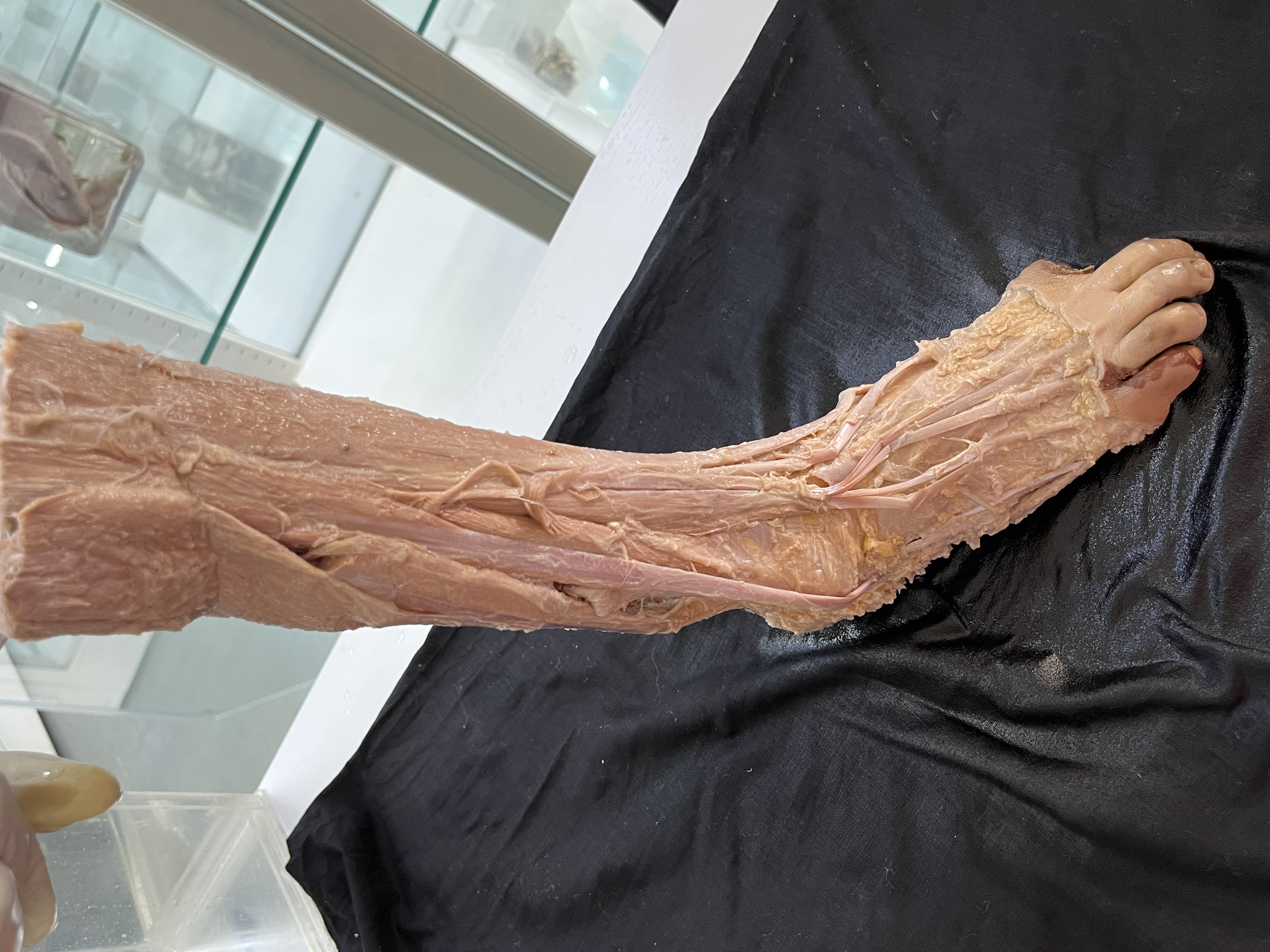
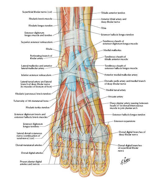
DORSUM OF FOOT
Home
⟶
DORSUM OF FOOT
Specimen Image
Rack Number
RACK 4 ABDOMEN AND LOWER LIMB
Specimen Number
18
↑