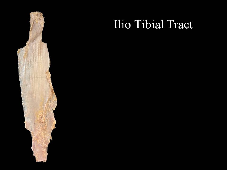
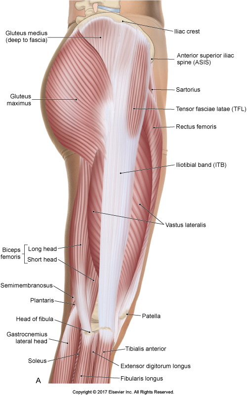
The iliotibial band is one of the most common running injuries. It is considered a non-traumatic overuse injury and is often concomitant with underlying weakness of hip abductor muscles. For more see Iliotibial Band Syndrome. Clinical examination testing for ITB dysfunction is best elicited utilizing the Ober Test, see here
External snapping hip syndrome is another ITB pathology. Snapping Hip Syndrome is a condition that is characterized by a snapping sensation, and/or audible “snap” or “click” noise, in or around the hip when it is in motion.
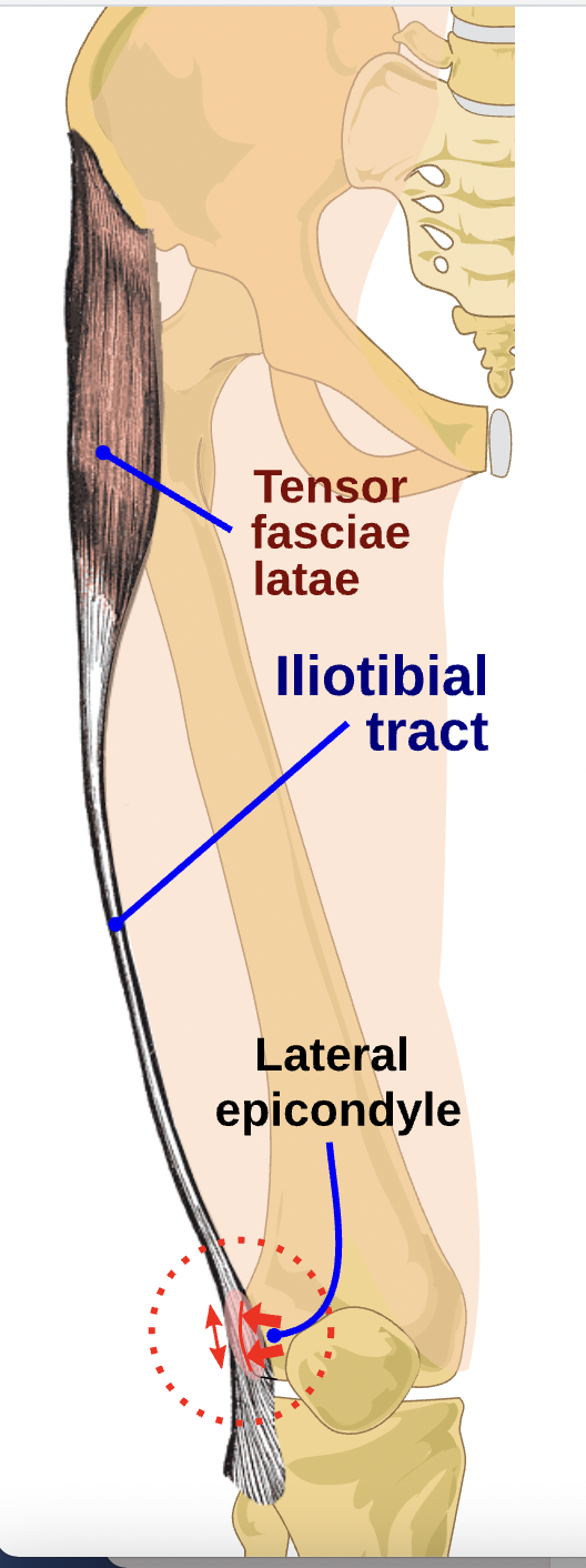
It has been proposed that a tight iliotibial band (ITB) through its attachment of the lateral retinaculum into the patella could cause lateral patella tracking, patella tilt and compression. This has implications in subjects presenting with patellofemoral pain syndrome (PFPS)