Skip to main content
NAMO
NAMO
NAMO
NAMO
NAMO
NAMO
NAMO
x
Search
Search
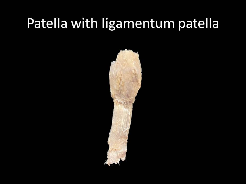
Patella with Ligamentum patellae
Home
⟶
Patella with Ligamentum patellae
Specimen Image
Rack Number
RACK 3 THORAX AND UPPER LIMB
Specimen Number
43
↑