Skip to main content
NAMO
NAMO
NAMO
NAMO
NAMO
NAMO
NAMO
x
Search
Search
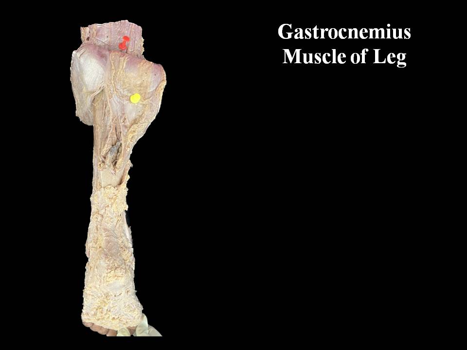
Gastrocnemius muscle of Leg
Home
⟶
Gastrocnemius muscle of Leg
Specimen Image
Rack Number
RACK 3 THORAX AND UPPER LIMB
Specimen Number
42
↑