Skip to main content
NAMO
NAMO
NAMO
NAMO
NAMO
NAMO
NAMO
x
Search
Search
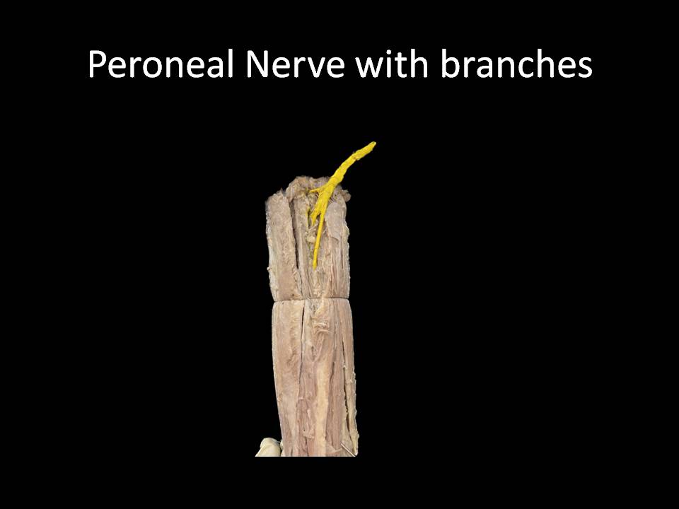
Peroneal Nerve with Branches
Home
⟶
Peroneal Nerve with Branches
Specimen Image
Rack Number
RACK 3 THORAX AND UPPER LIMB
Specimen Number
40
↑