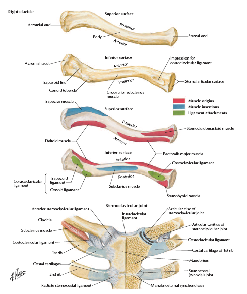
Fractures of the clavicle are common injuries accounting for between 2.6 and 4% of adult fractures and 35% of injuries to the shoulder girdle [1-3]. Early reports of clavicle fractures date back to Hippocrates [4], who noted that “when a fractured clavicle is fairly broken across it is more easily treated, but when broken obliquely it is more difficult to manage”. Clavicular fractures are most common in younger patients with incidence greatest in the second and third decades. The prevalence of fractures to the clavicle has been seen to decrease with every decade, after a patient is 20 years of age. However, the ratio of female to male increases with age. The aetiology of clavicle injuries in young adults and children is most commonly an RTA, sports injury and, to a lesser extent, a fall. However, falls represent their most frequent cause among the elderly [3]. Clavicle injuries can be grossly divided into three distinct anatomical sites; the medial clavicle, shaft and lateral end. Mid-shaft clavicle [1, 3] fractures are most common, with an incidence of up to 82% of all clavicle fractures. Medial and lateral end fractures account for approximately 18 and 2% respectively [3]. The location and pattern of injury are of considerable importance when formulating a management plan.
A number of classification systems have been described for the classification of clavicle fractures [1, 5-7]. Allman [5] divided clavicle fractures by anatomical site into 3 groups; group 1 being fractures to the middle third, group 2 being fractures distal to the coraco-clavicular ligament, and Group 3 relating to fractures of the proximal third of the clavicle [5]. Neer [6] went further and subdivided lateral third fractures into three groups; undisplaced, displaced, and intraarticular. The displaced types were then divided into 2a or 2b, depending on the presence of injury to the coracoclavicular (CC) ligaments [6]. Thus a type 2a injury represents a fracture medial to both conoid and trapzezoid elements of the CC ligaments, with the shaft displacing superior relative to the lateral end. A type 2b injury represents a fracture of the lateral end of the clavicle, with disruption of the conoid portion of the CC ligament [6]. Robinson [1] was the first to describe clavicle fractures in relation to their displacement and degree of comminution, via the Edinburgh classification. He then used his parameters to predict the risk of non-union, in such fractures, with good affect [7]. The Edinburgh classification system has been shown to provide more reliable prognostic information in middle third fractures, in comparison to other classification systems.
