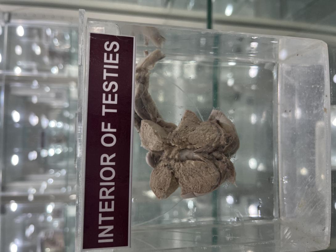
The testes — also called testicles — are two oval-shaped organs in the male reproductive system. They’re contained in a sac of skin called the scrotum. The scrotum hangs outside the body in the front of the pelvic region near the upper thighs.
Structures within the testes are important for the production and storage of sperm until they’re mature enough for ejaculation. The testes also produce a hormone called testosterone. This hormone is responsible for sex drive, fertility, and the development of muscle and bone mass.
Anatomy and function of testes
The main function of the testes is producing and storing sperm. They’re also crucial for creating testosterone and other male hormones called androgens.
Testes get their ovular shape from tissues known as lobules. Lobules are made up of coiled tubes surrounded by dense connective tissues.
Seminiferous tubules
Seminiferous tubules are coiled tubes that make up most of each testis. The cells and tissues in the tubules are responsible for spermatogenesis, which is the process of creating sperm.
These tubules are lined with a layer of tissue called the epithelium. This layer is made up of Sertoli cells that aid in the production of hormones that generate sperm. Among the Sertoli cells are spermatogenic cells that divide and become spermatozoa, or sperm cells.
The tissues next to the tubules are called Leydig cells. These cells produce male hormones, such as testosterone and other androgens.
Rete testis
After sperm is created in the seminiferous tubules, sperm cells travel toward the epididymis through the rete testis. The rete testis helps to mix sperm cells around in the fluid secreted by Sertoli cells. The body reabsorbs this fluid as sperm cells travel from the seminiferous tubules to the epididymis.
Before sperm can get to the epididymis, they can’t move. Millions of tiny projections in the rete testis, known as microvilli, help move sperm along to the efferent tubules.
Efferent ducts
The efferent ducts are a series of tubes that join the rete testis to the epididymis. The epididymis stores sperm cells until they’re mature and ready for ejaculation.
These ducts are lined with hair-like projections called cilia. Along with a layer of smooth muscle, cilia help move the sperm into the epididymis.
The efferent ducts also absorb most of the fluid that helps to move sperm cells. This results in a higher concentration of sperm in ejaculate fluid.
Tunica: Vasculosa, albuginea, and vaginalis
The testes are surrounded by several layers of tissue. They are the:
- tunica vasculosa
- tunica albuginea
- tunica vaginalis
Tunica vasculosa is the first thin layer of blood vessels. This layer shields the tubular interior of each testicle from further layers of tissue around the outer testicle.
The next layer is called the tunica albuginea. It’s a thick, protective layer made of densely packed fibers that further protect the testes.
The outermost layers of tissue are called the tunica vaginalis. The tunica vaginalis consists of three layers:
- Visceral layer. This layer surrounds the tunica albuginea that shields the seminiferous tubules.
- Cavum vaginale. This layer is an empty space between the visceral layer and the outermost layer of the tunica vaginalis.
- Parietal layer. This layer is the outermost protective layer that surrounds almost the entire testicular structure.

