The small intestine is an organ located within the gastrointestinal tract. It is approximately 6.5m in the average person and assists in the digestion and absorption of ingested food.
It extends from the pylorus of the stomach to the ileocaecal junction, where it meets the large intestine at the ileocaecal valve. Anatomically, the small bowel can be divided into three parts: the duodenum, jejunum, and ileum.
In this article, we shall examine the anatomy of the small intestine – its structure, neurovascular supply, and clinical correlations.
By TeachMeSeries Ltd (2023)
Fig 1 – The anatomical divisions of the small intestine.
Fig 1 – The anatomical divisions of the small intestine.
The Duodenum
The most proximal portion of the small intestine is the duodenum. Its name is derived from the Latin ‘duodenum digitorum’, meaning twelve fingers length. It runs from the pylorus of the stomach to the duodenojejunal junction.
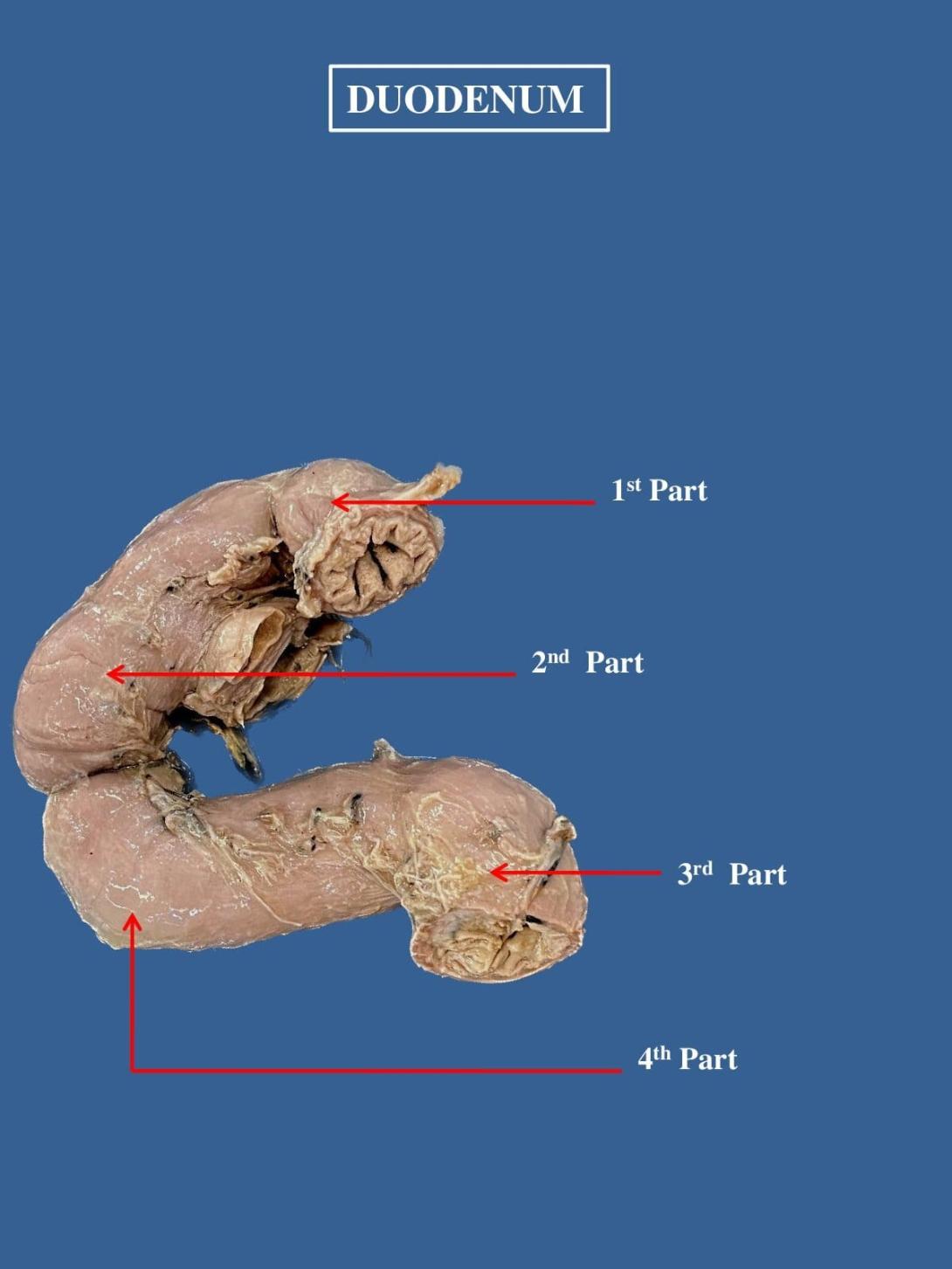
The duodenum can be divided into four parts: superior, descending, inferior and ascending. Together these parts form a ‘C’ shape, that is around 25cm long, and which wraps around the head of the pancreas.
D1 – Superior (Spinal level L1)
The first section of the duodenum is known as ‘the cap’. It ascends upwards from the pylorus of the stomach, and is connected to the liver by the hepatoduodenal ligament. This area is most common site of duodenal ulceration.
The initial 3cm of the superior duodenum is covered anteriorly and posteriorly by visceral peritoneum, with the remainder retroperitoneal (only covered anteriorly).
D2 – Descending (L1-L3)
The descending portion curves inferiorly around the head of the pancreas. It lies posteriorly to the transverse colon, and anterior to the right kidney.
Internally, the descending duodenum is marked by the major duodenal papilla – the opening at which bile and pancreatic secretions to enter from the ampulla of Vater (hepatopancreatic ampulla).
D3 – Inferior (L3)
The inferior duodenum travels laterally to the left, crossing over the inferior vena cava and aorta. It is located inferiorly to the pancreas, and posteriorly to the superior mesenteric artery and vein.
D4 – Ascending (L3-L2)
After the duodenum crosses the aorta, it ascends and curves anteriorly to join the jejunum at a sharp turn known as the duodenojejunal flexure.
Located at the duodenojejunal junction is a slip of muscle called the suspensory muscle of the duodenum. Contraction of this muscle widens the angle of the flexure, and aids movement of the intestinal contents into the jejunum.
By TeachMeSeries Ltd (2023)

