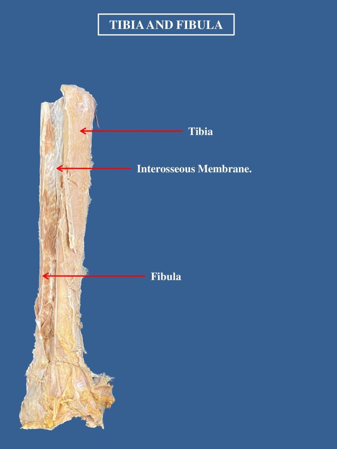
The proximal and distal tibiofibular joints refer to two articulations between the tibia and fibula of the leg. These joints have minimal function in terms of movement but play a greater role in stability and weight-bearing.
In this article, we shall look at the anatomy of the proximal and distal tibiofibular joints – their structures, neurovascular supply and clinical relevance.
Adapted from work by OpenStax College [CC BY 3.0], via Wikimedia Commons
Fig 1 – Anterior view of the right proximal and distal tibiofibular joints.
Proximal Tibiofibular Joint
Articulating Surfaces
The proximal tibiofibular joint is formed by an articulation between the head of the fibula and the lateral condyle of the tibia.
It is a plane type synovial joint; where the bones to glide over one another to create movement.
Supporting Structures
The articular surfaces of the proximal tibiofibular joint are lined with hyaline cartilage and contained within a joint capsule.
The joint capsule receives additional support from:
- Anterior and posterior superior tibiofibular ligaments – span between the fibular head and lateral tibial condyle
- Lateral collateral ligament of the knee joint
- Biceps femoris – provides reinforcement as it inserts onto the fibular head.
Neurovascular Supply
The arterial supply to the proximal tibiofibular joint is via the inferior genicular arteries and the anterior tibial recurrent arteries.
The joint is innervated by branches of the common fibular nerve and the nerve to the popliteus (a branch of the tibial nerve).

