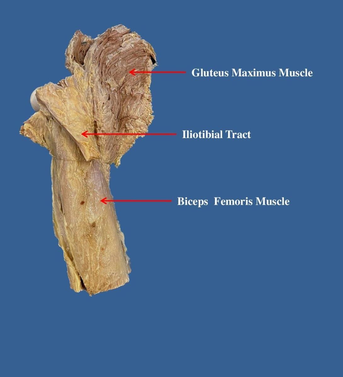
The gluteal region is an anatomical area located posteriorly to the pelvic girdle, at the proximal end of the femur. The muscles in this region move the lower limb at the hip joint.
The muscles of the gluteal region can be broadly divided into two groups:
- Superficial abductors and extenders – group of large muscles that abduct and extend the femur. Includes the gluteus maximus, gluteus medius, gluteus minimus and tensor fascia lata.
- Deep lateral rotators – group of smaller muscles that mainly act to laterally rotate the femur. Includes the quadratus femoris, piriformis, gemellus superior, gemellus inferior and obturator internus.
The arterial supply to these muscles is mostly via the superior and inferior gluteal arteries – branches of the internal iliac artery. Venous drainage follows the arterial supply.
In this article, we shall examine the two groups of gluteal muscles – their attachments, innervations and actions. We shall also look at the clinical consequence of gluteal muscle disorders.
The Superficial Muscles
The superficial muscles in the gluteal region consist of the three glutei and the tensor fascia lata. They mainly act to abduct and extend the lower limb at the hip joint.
Clinical Relevance: Damage to the Superior Gluteal Nerve
The superior gluteal nerve innervates the gluteus medius and the gluteus minimus. These muscles have an important role in stabilising the pelvis during locomotion. In the standing position, the gluteus minimus and medius contract when the contralateral leg is raised, preventing the pelvis from dropping on that side.
If the superior gluteal nerve is damaged, the previously described muscles are paralysed – and the pelvis becomes unsteady. A characteristic finding of gluteal muscle weakness is the Trendelenburg sign.
Trendelenburg Sign
The Trendelenburg sign is produced when the patient is asked to stand unassisted on each leg in turn. In a positive sign, pelvic drop will occur on the unsupported leg. Pelvic drop can be recognised by observing the level of the iliac crests on both sides.
For example, if the left gluteal muscles are weak, the right side of the pelvis will drop when the patient stands on their left leg (and the right leg is unsupported).

