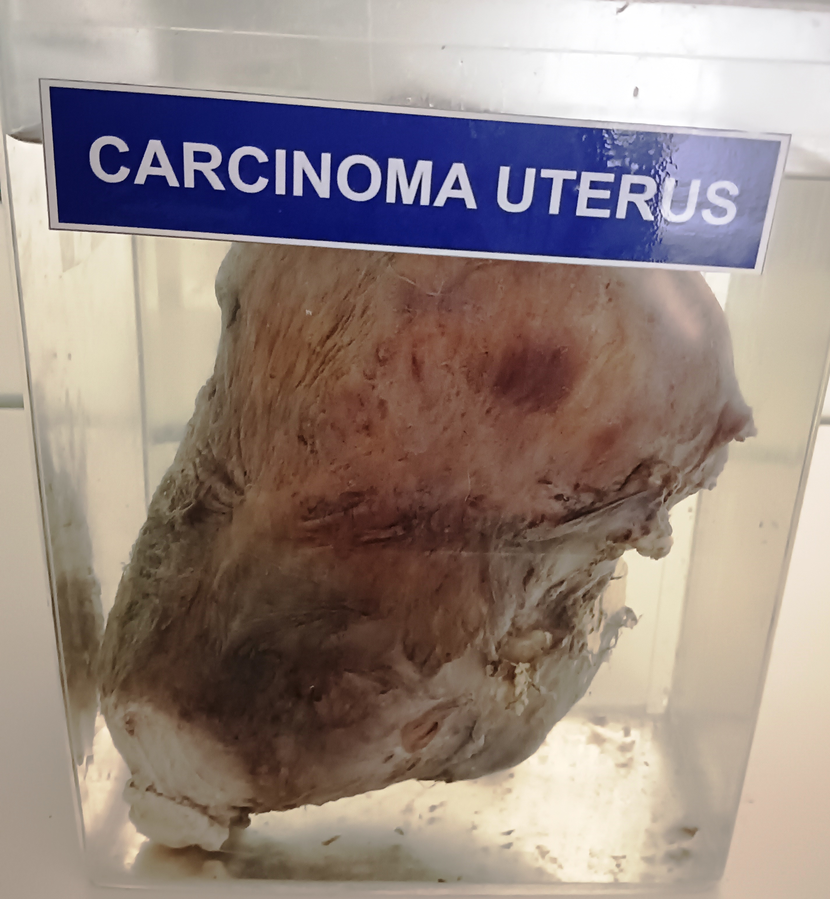
NAMO
NAMO
NAMO
NAMO
NAMO
NAMO
NAMO
Specimen Image

Key Points

  • Uterine fibroids (also known as myomas or leiomyomas) are benign monoclonal neoplasms of the myometrium that represent the most common gynecologic tumor. They may be single or multiple, of variable size
    • They occur more often in Black patients; incidence and symptoms increase until menopause, at which point existing lesions and symptoms regress
  • Many patients are asymptomatic. When symptoms do occur, they may be related to excess bleeding or to size and location of lesions and may include dysmenorrhea, menorrhagia, metrorrhagia, dyspareunia, bladder dysfunction (urinary frequency, incontinence, or retention), constipation, and pelvic pain and/or fullness
    • Reproductive effects may include infertility, recurrent miscarriage, premature birth, abnormal presentation, placenta abruptio, and intrauterine growth restriction
  • Because treatment is indicated only for symptomatic fibroids and because malignant transformation is not thought to occur, there are no standard recommendations for monitoring asymptomatic fibroids
  • When treatment of symptomatic fibroids is desired, tailor the approach to the nature of symptoms (ie, bleeding, bulk, or both); number, location, and size of fibroids; and patient's reproductive plans
    • Hysterectomy is the definitive treatment, but it carries inherent surgical risks and is not suitable for patients who wish to preserve childbearing potential
    • Less invasive measures include myomectomy, uterine artery embolization, ablation with high-frequency ultrasonography, and other less common techniques. These measures are generally quite effective in relieving symptoms caused by bleeding or size while retaining fertility
    • Pharmacologic therapy is indicated for urgent control of severe bleeding, to reduce fibroid size and correct anemia before surgical intervention, and as a bridge to anticipated menopause and decline in fibroid size
      • Heavy menstrual bleeding can be controlled with NSAIDs, tranexamic acid, hormonal contraceptives, or gonadotropin-releasing hormone antagonists, but these have little or no effect on fibroid size
      • Gonadotropin-releasing hormone agonists and selective progesterone receptor modulators can be used to reduce size and bulk as well as to diminish bleeding
  • Size and symptoms of fibroids regress after menopause. Whether malignant transformation of fibroids contributes to development of leiomyosarcoma is not known with certainty; however, malignant degeneration of fibromas is exceedingly rare

Pitfalls

  • Most pharmacologic treatments that are effective in reducing fibroid size are limited to short-term use (eg, 6 months) owing to menopausal symptoms and adverse effects on bone density
  • Morcellation of the uterus or uterine myomas carries risk of dissemination of an occult malignancy 1

Terminology

Clinical Clarification

  • Uterine fibroids (also known as myomas or leiomyomas) are benign monoclonal neoplasms of the myometrium that represent the most common gynecologic tumor
    • May be single or multiple, of variable size
    • May be asymptomatic or may cause menstrual abnormalities, anemia, and pelvic pain
    • Occur in females of reproductive age and regress after menopause

Classification

  • Commonly classified based on the location in the uterus 2
    • Submucosal: arising just beneath the endometrium
    • Intramural: arising within the uterine wall
    • Subserosal: arising from the serosal surface
    • Transmural: extending from the endometrium to the serosal surface
  • Most detailed classification is the International Federation of Gynecology and Obstetrics scheme, based on location and structure 2
    • 0: intracavitary, pedunculated
    • 1: submucosal (less than 50% intramural)
    • 2: submucosal (50% or more intramural)
    • 3: intramural with endometrial contact
    • 4: intramural
    • 5: subserosal (50% or more intramural)
    • 6: subserosal (less than 50% intramural)
    • 7: subserosal, pedunculated
    • 8: other (eg, cervical)

Diagnosis

Clinical Presentation

History

  • Many patients are asymptomatic
  • In most symptomatic patients, nature of the concern depends on size and location of lesions and may include:
    • Irregular (metrorrhagia) and/or heavy (menorrhagia) menstrual bleeding
    • Dysmenorrhea
    • Noncyclic pelvic pain
    • Dyspareunia
    • Pelvic and/or abdominal fullness
    • Urinary frequency, incontinence, or retention
    • Constipation
    • Symptoms associated with anemia (eg, fatigue)
  • Other symptoms may be related to fertility or pregnancy:
    • Infertility
    • Recurrent miscarriage

Physical examination

  • Bimanual examination may show a distended uterus (sometimes equivalent to third trimester of pregnancy) with palpable, knoblike irregularities
  • Occasionally, a fibroid may be seen protruding through the cervix on speculum examination
  • Patients with a long history of menorrhagia may exhibit signs of anemia such as pallor with pale conjunctivae and nailbeds

Causes and Risk Factors

Causes

  • Monoclonal proliferation, most likely resulting from heightened responsiveness to estrogen and progesterone in a genetically susceptible myometrial cell 3

Risk factors and/or associations

Age
  • Risk increases with age from time of menarche until menopause, at which point it declines significantly
Genetics
  • Several mutations that appear to increase sensitivity to estrogen and progesterone have been identified: 4
    • MED12 mutations, resulting in alterations in cell signaling and increased cell proliferation; found in approximately 70% of fibroids 4
    • Deletions within Xq22.3 locus of COL4A5 and COL4A6 genes, which result in enhanced cell proliferation 5
    • Inactivation of the fumarate hydratase gene; occurs in a small percentage of sporadic fibroids and in the rare hereditary syndrome of leiomyomatosis and renal cell cancer 5 6
Ethnicity/race
  • More common in Black patients 3 7
    • Lifetime risk is approximately 70% for White females and over 80% for Black females
    • Tumors tend to be larger, more numerous, and more symptomatic, and develop at a younger age in Black patients than in White patients
    • Clinically significant fibroids (defined as a uterine size equivalent to 9 weeks or more of gestation, at least 1 tumor bigger than 4 cm, or 1 submucosal lesion) occur twice as often in Black patients as in White patients
Other risk factors/associations
  • Nulliparity increases risk; multiparity and older age at first full-term pregnancy appear to reduce risk 3
  • Increased risk has been associated with BMI defined as overweight, but some studies show lower risk with BMI defined as obese compared with overweight 3
  • Risk may be reduced by physical exercise, although the parameters that provide benefit are not clearly defined 3
  • Use of depot medroxyprogesterone acetate reduces risk, which declines progressively with longer duration of use 3
  • Roles of dietary and other factors are the subject of ongoing study
    • Vitamin D supplementation has been associated with a decrease in risk 8

Diagnostic Procedures

  • Image removed.

    Submucosal fibroid with saline infusion sonography.

    From Mitchell C et al: Role of ultrasound in evaluating female infertility. In: Hagen-Ansert SL, ed: Textbook of Diagnostic Sonography. 8th ed. Elsevier, Inc.; 2018:1151-8, Figure 46-6

  • Image removed.

    (A) 2-dimensional long-axis image of the uterus taken during a sonohysterogram showing a posterior partly submucous fibroid. - (B) 3-dimensional coronal view of the same uterus showing that the fibroid involves the left lateral and cornual aspect of the cavity.

    From Benacerraf BR: Three-dimensional volume imaging in gynecology. Obstet Gynecol Clin North Am. 46(4): 755-81, 2019, Figure 34

  • Image removed.

    Uterine fibroid on transvaginal ultrasonogram. - A hypoechoic mass with areas of shadowing is seen within the body of the retroverted uterus, the typical sonographic appearance of a uterine fibroid. This intramural fibroid causes a slight bulge in the external uterine contour.

    From Zagoria RJ et al: The female genital tract. In: Genitourinary Imaging: The Requisites. 3rd ed. Elsevier, Inc.; 2016:248-303, Figure 7-50

  • Image removed.

    Multiple small leiomyomas in the posterior uterine cavity wall as observed during endometrial resection.

    From Kumar A et al: Localized subendometrial leiomyomatosis at hysteroscopy. J Minim Invasive Gynecol. 19(3):284-28, 2012, Figure 1


Primary diagnostic tools

  • History and physical examination findings may suggest the diagnosis in many patients 8
    • Exclude pregnancy in patients of childbearing age
  • Ultrasonography is the initial diagnostic test of choice 9 10 11
  • Saline infusion sonohysterography may define the endometrium and cavity more clearly if conventional ultrasonography suggests a submucosal or pedunculated intracavitary lesion 9 12
  • Conventional hysteroscopy can differentiate pedunculated intracavitary fibroids from endometrial polyps or provide clarity if there is a suspicion of malignancy requiring an endometrial biopsy 9
  • MRI may be indicated to provide more detail regarding the number and depth of fibroids detected by ultrasonography, if ultrasonography is nondiagnostic, or in the setting of abnormal vaginal bleeding if an intracavitary mass is suspected and hysteroscopy is not feasible 9 12
  • Recommended to measure hemoglobin and hematocrit levels in patients who report heavy bleeding 9


Laboratory

  •  
    •  


Imaging

  •  
    •  
    •  
  •  
    •  
    •  
    •  
    •  
  •  
    •  
    •  
    •  


Procedures


Differential Diagnosis

Most common

  • Symptoms predominately menstrual (ie, heavy and/or irregular bleeding, dysmenorrhea):
    • Menopausal transition (Related: Perimenopause and Menopause)
      • Like fibroids, may cause irregular or heavy bleeding in patients in their late 40s through early 50s
      • Unlike fibroids, menopausal transition may be accompanied by symptoms associated with hypoestrogenism (eg, hot flashes)
      • Menopause is characterized by follicle-stimulating hormone level higher than 30 to 40 milliunits/L
      • Owing to increasing prevalence of fibroids during the fifth decade, perimenopausal symptoms may converge with those caused by fibroids; however, symptoms caused by fibroids tend to subside as menopause progresses
    • Endometrial polyps
      • Common endometrial or endocervical epithelial proliferations consisting of variable vascular, glandular, fibromuscular, and connective tissues
      • Like fibroids, may present with abnormal bleeding, and ultrasonography may show intracavitary mass
      • May be differentiated from fibroids by serial ultrasonography showing typical evolution during the course of the menstrual cycle or, more commonly, by sonohysterography or hysteroscopy
    • Adenomyosis
      • Disruptive pockets of endometrial glands and stroma internal to myometrium
      • Like fibroids, occur most commonly in patients aged 35 to 50 years and may be associated with dysmenorrhea, noncyclic pelvic pain, and menorrhagia
      • Differentiated from fibroids by ultrasonography or MRI; definitive diagnosis is made on histologic examination after hysterectomy
    • Endometriosis
      • Inflammatory disease in which ectopic endometrial-like tissue forms lesions outside the uterus
      • Like fibroids, may present with dysmenorrhea, infertility, menorrhagia, long-term pelvic discomfort, and dyspareunia
      • Occurs primarily in younger patients
      • Diagnosis usually can be made by clinical presentation and imaging; laparoscopy and biopsy provide a definitive diagnosis of endometriosis if imaging is not diagnostic
    • Endometrial hyperplasia or carcinoma (Related: Endometrial Hyperplasia)
      • Endometrial hyperplasia is a thickening of the endometrium attributable to increased proliferation of endometrial glands relative to stroma; may undergo malignant transformation to endometrial carcinoma
      • Like fibroids, typically manifests with abnormal uterine bleeding and pelvic pain
      • Endometrial carcinoma is uncommon, particularly in younger patients; however, clinical presentation may be indistinguishable from fibroids (Related: Endometrial Carcinoma)
      • Differentiation suggested by imaging findings; endometrial biopsy confirms diagnosis
  • Symptoms predominately caused by bulk and location (ie, urinary symptoms, constipation, pelvic and/or abdominal fullness)
    • Pregnancy
      • Like fibroids, may be associated with pelvic and/or abdominal fullness, urinary or bowel symptoms, and uterine enlargement
      • Bluish discoloration of cervix and vagina and cervical softening suggest pregnancy
      • Differentiation is based on positive pregnancy test result and ultrasonography findings
      • Pregnancy and fibroids may occur concomitantly, and presence of fibroids may complicate pregnancy
    • Mass lesions caused by pelvic organ malignancy
      • Bulky lesions of colon, uterus, bladder, ovaries, cervix, fallopian tubes, or kidneys may result in feeling of fullness, distention, and pelvic pain, and may cause dysfunction of bowel and/or bladder
      • Pain may be progressively severe
      • Differentiation is made by imaging

Treatment

Goals

  • Relieve symptoms caused by excessive bleeding or size of fibroids
  • Prevent further growth
  • Restore fertility

Disposition

Admission criteria

  • Severe bleeding requiring transfusion and urgent medical or surgical therapy
  • Required for some elective treatment modalities (eg, hysterectomy, abdominal myomectomy, uterine artery embolization)

Recommendations for specialist referral

  • Refer to an obstetrician/gynecologist for selection and management of appropriate pharmacotherapy or surgical intervention (myomectomy or hysterectomy)
  • Refer to an interventional radiologist for uterine artery embolization or MR–guided focused ultrasonography

Treatment Options

Therapy is generally indicated only for symptomatic fibroids, including infertility when they are considered to play a significant role 8

For many patients of reproductive age, medical management should be considered before moving to other, more invasive treatment options, including: 17

  • Patients with uterine fibroids who are symptomatic with heavy uterine bleeding or bulk symptoms (eg, pressure, pain, fullness, bladder, or bowel symptoms) and want to preserve fertility 17
  • Patients with uterine fibroids who are symptomatic with heavy uterine bleeding or bulk symptoms (eg, pressure, pain, fullness, bowel, or bladder symptoms) and do not want to preserve fertility 17
  • Patients with both uterine fibroids and adenomyosis who are symptomatic with heavy uterine bleeding or bulk symptoms (eg, pressure, pain, fullness, bladder, or bowel symptoms) and do not want to preserve fertility 17
  • Patients with pedunculated submucosal uterine fibroids who are symptomatic with heavy uterine bleeding 17

Selection of treatment modality depends on the nature of symptoms (ie, bleeding versus pain or bulk); location, size, and number of fibroids; and the patient's age and reproductive plans 18 19 20

  • Hysterectomy is the only permanent curative treatment, but it carries operative risks and eliminates childbearing potential
    • Up to 90% of patients experience at least moderate improvement in symptoms 21
    • Symptom improvement may not be immediate but is reported in approximately 80% of patients by 6 to 12 months 22
  • If a patient chooses to preserve childbearing potential or retain their uterus, consider alternatives to hysterectomy
    • Surgical myomectomy, uterine artery embolization, and ablation with MR-guided ultrasonography offer potential for more durable relief in patients who choose not to have hysterectomy but whose symptoms are attributed to large fibroids 22
      • Myomectomy may be done via open abdominal approach, laparoscopically, or hysteroscopically depending on the size, location, and type of fibroid. Laparoscopic myomectomy results in fewer complications and shorter recovery than open myomectomy, but not all patients are suitable candidates; hysteroscopic myomectomy is an option for submucosal leiomyomas 23 24
        • A 2020 randomized trial suggested that, among patients with symptomatic uterine fibroids, those undergoing myomectomy experienced better fibroid-related quality of life at 2 years than those undergoing uterine artery embolization 25
        • Approximately 20% of patients who are treated by myomectomy require a second procedure owing to recurrent symptoms
        • A Cochrane review of myomectomy in treatment of infertility found insufficient evidence to assess the efficacy of myomectomy for this indication. Comparison between various myomectomy techniques did not indicate any method was superior in improving rates of live birth, preterm delivery, clinical pregnancy, ongoing pregnancy, miscarriage, or cesarean delivery 23 26
      • Uterine artery embolization is a minimally invasive option with high initial success rates (82%-90% for menorrhagia and 77%-86% for dysmenorrhea and symptoms due to size), but approximately 10% of patients require intervention within several years owing to recurrent symptoms 22
      • Ablation with MR-guided ultrasonography improves symptoms in approximately 80% of patients by 6 to 12 months and has a low rate of adverse effects; however, long-term results are uncertain 22 27
        • Reported failure rates are approximately 4%, 11%, and 24% at 6-month, 12-month, and 5-year follow-ups, respectively 27
      • In parts of Europe, transcervical ultrasonography–guided radiofrequency ablation has been used to treat symptomatic fibroids 28
      • Ultrasonography-guided, high-intensity-focused ultrasonography ablation is a commonly used modality in China 29 30
    • Less common procedures include laparoscopic or vaginal ligation of the uterine arteries and laparoscopic cryomyolysis or thermocoagulation 9
    • Pharmacologic treatment options include medications that treat only abnormal bleeding symptoms (gonadotropin-releasing hormone antagonists, levonorgestrel-releasing IUDs (intrauterine devices), hormonal contraceptives, tranexamic acid) and medications that reduce both bleeding and leiomyoma size (gonadotropin-releasing hormone agonists and selective progesterone receptor modulators) 7
      • Currently available pharmacologic treatment options primarily provide symptomatic treatment; further research on potential long-term treatments is needed 31
      • Heavy menstrual bleeding can be controlled with NSAIDs, tranexamic acid, or hormonal contraceptives; these have little or no effect on fibroid size (Related: Abnormal Uterine Bleeding in Females of Reproductive Age)8
        • Levonorgestrel-releasing IUDs decrease heavy menstrual bleeding in patients both with and without leiomyomas; however, there is insufficient evidence to support their use in treatment of other uterine leiomyoma symptoms 7 32
        • Progestin-only or combination estrogen-progesterone contraceptive agents are also reasonable options 7
          • However, there is evidence that progesterone and progestogens play a role in the pathogenesis of uterine myomas and treatment may cause an increase in fibroid size 33
        • Tranexamic acid is an effective treatment for patients with heavy menstrual bleeding, including that associated with leiomyomas 7
        • Gonadotropin-releasing hormone antagonists
          • Elagolix and relugolix are both FDA approved for treatment of heavy menstrual bleeding associated with uterine leiomyomas 7 34 35 36 37
          • Preferred treatment option owing to efficacy and relatively mild adverse effects 38
          • Can be used for up to 2 years, in conjunction with hormonal add-back therapy 7
          • Hormonal add-back therapy is indicated to offset the hypoestrogenic effects including hot flashes, increased serum lipid levels, and bone mineral density loss 39 40
      • Hormonal agents that are effective at reducing both size and bulk of fibroids and bleeding are generally limited to short-term use; fibroid growth and symptoms resume several months after discontinuation 4
        • Indications include:
          • For urgent control of severe bleeding
          • To reduce fibroid size and correct anemia before interventional procedures or surgical management
          • As a bridge to anticipated menopause and decline in fibroid size
        • Options include: 4
          • Gonadotropin-releasing hormone agonists (eg, leuprolide, goserelin)
            • Primarily used as a short-term bridging treatment
              • Treatment is typically limited to 6 months without add-back therapy and 12 months with add-back therapy
            • Reduces leiomyoma size and overall size of the uterus, heavy bleeding, and dysmenorrhea 7
              • When used preoperatively, reduction in uterine volume may then allow the use of a minimally invasive surgical route or a smaller incision 7
            • Associated with an initial flare of symptoms followed by reduction or absence of menstrual blood flow; often attended by symptoms of menopause (eg, hot flashes, vaginal dryness)
            • Add-back therapy can be used to reduce hypoestrogenic effects
              • Add-back regimens include progesterone, estrogen, combined estrogen and progesterone, tibolone (not available in the United States), ipriflavone, and raloxifene
              • A Cochrane review showed low to moderate evidence of a positive effect on preserving bone density with tibolone, raloxifene, estriol, and ipriflavone and a reduction in vasomotor symptoms with medroxyprogesterone acetate and tibolone; larger uterine sizes were associated with use of conjugated estrogens, medroxyprogesterone, and tibolone 41
          • Selective progesterone receptor modulators (eg, mifepristone, ulipristal)
            • Mifepristone and ulipristal acetate are effective in treatment of heavy bleeding and uterine enlargement associated with uterine leiomyomas but are not currently approved in the United States for the treatment of leiomyomas 7
            • Prolonged intermittent administration of these agents can be used to treat fibroid-related symptoms and is generally effective, safe, and well tolerated 42 43
            • Effective in reducing bleeding and fibroid size, with fewer vasoactive adverse effects and less effect on bone density than gonadotropin-releasing hormone analogues 44 45
            • Ulipristal is associated with effects that last longer than those of other pharmacologic agents (up to 6 months 46 after drug discontinuation), and courses may be repeated 13 46
              • Withdrawn from market in Canada and restricted use in Europe owing to reported cases of serious liver injury, including some requiring transplantation 47 48
          • Other agents that may have potential roles in treatment of fibroids include androgens (eg, danazol), aromatase inhibitors (eg, letrozole), and selective estrogen receptor modulators (eg, raloxifene)
            • Use of aromatase inhibitors has been proposed to reduce bulk; individual studies have shown significant treatment response, but a Cochrane review concluded that evidence was insufficient to support a recommendation 45 49
      • Several drugs and dietary components are under study regarding their role in uterine fibroid development and treatment; however, further research evidence is needed 31

Drug therapy

  • Agents to control menorrhagia
    • Antifibrinolytic agent
      • Tranexamic acid
        • Tranexamic Acid Oral tablet; Adults: 1,300 mg PO 3 times daily for up to 5 days during monthly menstruation.
    • NSAIDs
      • Naproxen
        • Naproxen Sodium Oral tablet; Adults: 550 mg PO once, then 550 mg PO every 12 hours as needed.
      • Mefenamic acid
        • Mefenamic Acid Oral capsule; Adults: 500 mg PO once, then 250 mg PO every 6 hours as needed.
    • Hormonal contraceptives
      • Levonorgestrel
        • Levonorgestrel Vaginal insert; Adults: 52 mg intrauterine device (IUD) inserted into the uterus. Remove and replace the IUD after 5 years if continued use is needed.
      • Levonorgestrel/ethinyl estradiol
        • Levonorgestrel, Ethinyl Estradiol Oral tablet; Adults: 0.05 to 0.15 mg levonorgestrel/0.02 to 0.04 mg ethinyl estradiol PO once daily for 21 days, followed by 7 days of inert, inactive tablets as for routine contraception.
      • Medroxyprogesterone
        • Medroxyprogesterone Acetate Suspension for injection [Contraception]; Adults: 150 mg IM every 13 weeks.
      • Norethindrone/ethinyl estradiol
        • Ethinyl Estradiol, Norethindrone Oral tablet; Adults: 0.4 to 1 mg norethindrone/0.035 mg ethinyl estradiol PO once daily for 21 days, followed by 7 days of inert, inactive tablets as for routine contraception.
      • Norgestimate/ethinyl estradiol
        • Inert Oral tablet, Norgestimate, Ethinyl Estradiol Oral tablet; Adults: 0.18 to 0.25 mg norgestimate; 0.025 to 0.035 mg ethinyl estradiol PO once daily for 21 days, followed by 7 days of inert, inactive tablets as for routine contraception.
  • Agents to control menorrhagia and reduce fibroid size
    • Gonadotropin-releasing hormone agonists
      • Goserelin
        • Goserelin Acetate Implant; Adults: 3.6 mg subcutaneously every 28 days.
      • Leuprolide
        • 1-month depot injection
          • Leuprolide Acetate Suspension for injection [Endometriosis]; Adults: 3.75 mg IM once monthly for up to 3 months.
        • 3-month depot injection
          • Leuprolide Acetate Suspension for injection [Endometriosis]; Adults: 11.25 mg IM as a single dose, which provides a 3-month treatment course.
    • Gonadotropin-releasing hormone antagonists
      • Elagolix
        • Elagolix Oral capsule, Elagolix, Estradiol, Norethindrone Acetate Oral capsule; Adults: 300 mg elagolix/1 mg estradiol/0.5 mg norethindrone PO once daily in the morning and 300 mg elagolix PO once daily in the evening for up to 24 months.
      • Relugolix
        • Relugolix, Estradiol, Norethindrone Acetate Oral tablet; Adults: 40 mg relugolix/1 mg estradiol/0.5 mg norethindrone PO once daily for up to 24 months.
    • Selective progesterone receptor modulators
      • Mifepristone
        • Mifepristone Oral tablet [Pregnancy Termination]; Adults: 2.5 to 10 mg PO once daily.
    • Aromatase inhibitors
      • Letrozole
        • Letrozole Oral tablet; Adults: 2.5 mg PO once daily.
  • Add-back therapy for gonadotropin-releasing hormone agonist regimens longer than 6 months
    • Conjugated estrogens
      • Conjugated Estrogens Oral tablet; Adults: 0.625 mg PO once daily.
    • Medroxyprogesterone
      • Medroxyprogesterone Acetate Oral tablet; Adults: 5 to 20 mg PO once daily.
    • Norethindrone
      • Norethindrone Acetate Oral tablet; Adults: 2.5 to 5 mg PO once daily.
    • Raloxifene
      • Raloxifene Hydrochloride Oral tablet; Postmenopausal Adults: 60 mg PO once daily.

Nondrug and supportive care

Calcium supplementation is recommended for patients treated with gonadotropin-releasing hormone agonists

Procedures
Hysterectomy

General explanation

  • Surgical removal of the uterus, which may be performed via an abdominal or vaginal approach and may be open, laparoscopic, or vaginal with laparoscopic assistance

Indication

  • Symptomatic fibroids in patients who choose not to preserve childbearing potential

Contraindications

  • Pregnancy

Complications

  • Postoperative transfusion is required in approximately 2% of patients 45
  • Ureteral injury
  • Urinary incontinence
  • Vaginal prolapse
  • Morcellation has been used to enable hysterectomy through a smaller incision than standard; this has become controversial owing to the risk of disseminating malignant cells from an unsuspected leiomyosarcoma 45
    • Before considering morcellation of the uterus or myoma, evaluate for increased risk of uterine malignancy 1
    • Although the occurrence of lesions from disseminated malignant cells is very rare, guidelines recommend that patients be advised of the potential risks and benefits and alternatives to morcellation 1 18 45
Myomectomy

General explanation

  • Surgical removal of fibroids by hysteroscopic, laparoscopic, or open abdominal approach 8 45
  • A 2020 randomized trial suggested that, among patients with symptomatic uterine fibroids, those undergoing myomectomy experienced better fibroid-related quality of life at 2 years after surgery than those undergoing uterine artery embolization 25

Indication

  • May consider hysteroscopic myomectomy for symptomatic type 0 or 1 submucosal fibroids smaller than 3 cm 45
  • Laparoscopic myomectomy, with or without robotic assistance, may be appropriate when lesions are few and relatively small (especially when multiple) and easily reached by a laparoscope 9 45
  • Open abdominal myomectomy is preferred for deep or large fibroids or when more than 3 or 4 fibroids are to be removed 45

Contraindications

  • Laparoscopic myomectomy is contraindicated in patients with numerous, deep, or very large fibroids

Complications

  • Postoperative transfusion is required in 2% to 28% of patients 45
  • Intrauterine adhesions, which may impair subsequent fertility, may occur after hysteroscopic surgery; adnexal scarring may occur after open or laparoscopic myomectomy and may affect fertility 50
  • Uterine rupture in pregnancy may occur after laparoscopic myomectomy 45
  • Morcellation, mechanized slicing of fibroids into small pieces for laparoscopic extraction, has been associated with inadvertent seeding of the abdomen and pelvis with tissue fragments that generate parasitic leiomyomata; a thorough peritoneal lavage may avert this complication 9
    • Although the risk of inadvertent seeding is extremely low, the FDA issued a warning based on the description of at least 1 case in which morcellation of an unsuspected leiomyosarcoma resulted in dissemination of malignant cells with an ultimately fatal outcome
    • Before considering morcellation of the uterus or myoma, evaluate for increased risk of uterine malignancy 1
    • Guidelines recommend that when morcellation is anticipated, patients be advised of the risks and benefits and alternatives to morcellation 1 18 45
Uterine artery embolization 22

General explanation

  • Image-guided catheterization of uterine artery with injection of an embolic agent

Indication

  • Symptomatic uterine fibroids in patients who do not desire future pregnancy 45
  • Especially appropriate in patients with multiple or very large fibroids and in patients who are poor surgical candidates owing to other medical morbidities or physical conditions (eg, multiple previous abdominal or pelvic surgeries, extreme obesity) 51

Contraindications

  • Pregnancy
  • Active infection of uterus or adnexa

Complications

  • Severe pain caused by infarction of fibroids is the most common complication and may require hospitalization 45
  • A small percentage of patients develop ovarian dysfunction manifested by amenorrhea; this may result in permanent infertility 45
  • In individuals who become pregnant after the procedure, there is an increased risk of pregnancy complications (eg, miscarriage, preterm labor, abnormal presentation of the fetus, need for cesarean delivery) 45
Ablation with MR–guided focused ultrasonography 45 52

General explanation

  • Induce coagulation necrosis in uterine fibroids using MR to map and guide high-intensity ultrasonography

Indication

  • A single or a few symptomatic fibroids of moderate size (4-6 cm)

Contraindications

  • Uterine size equivalent to 24 weeks of gestation or larger
  • Pedunculated, nonenhancing, or heavily calcified fibroid
  • Significant abdominal scarring

Complications

  • Skin burns
  • Paresthesias of sciatic nerve

Special populations

  • Pregnant patients
    • Although fibroids have been associated with complications of pregnancy and delivery, myomectomy during pregnancy is not routinely recommended 53
    • Pharmacologic treatment of fibroids is contraindicated in pregnancy 4

Monitoring

  • Because treatment is indicated only for symptomatic fibroids and malignant transformation is not thought to occur, there are no standard recommendations for monitoring asymptomatic fibroids 8

Complications and Prognosis

Complications

  • Iron deficiency anemia 45
  • Infertility 45
    • Fibroids are associated with infertility in 10% of cases and are the only identified cause in approximately 5%
  • Recurrent miscarriage 54
  • There is a small increase in risk of some pregnancy-associated complications, primarily with submucosal fibroids: 50 55
    • Breech presentation
    • Placenta previa
    • Need for cesarean delivery
    • Placenta abruptio
    • Premature rupture of membranes
    • Premature birth
    • Intrauterine death with fetal growth restriction
  • Red degeneration (infarction) 20
  • Rarely, may result in life-threatening uterine hemorrhage 45
    • Urgent intervention is required: transfusion, antifibrinolytics (eg, tranexamic acid), IV estrogen, or high-dose oral contraceptives. Uterine artery embolization or hysterectomy may be required

Prognosis

  • Before menopause, pattern of fibroid growth is variable and unpredictable, but risk of symptoms increases until menopause
  • Fibroids (and associated symptoms) regress after menopause
  • The presence of fibroids in young patients does not appear to strongly influence ovarian reserve as estimated by antimüllerian hormone levels 56 57
  • Cause of leiomyosarcoma is unknown (ie, whether they arise de novo or from uterine fibromas); however, malignant degeneration of fibromas is exceedingly rare
Specimen Number
56