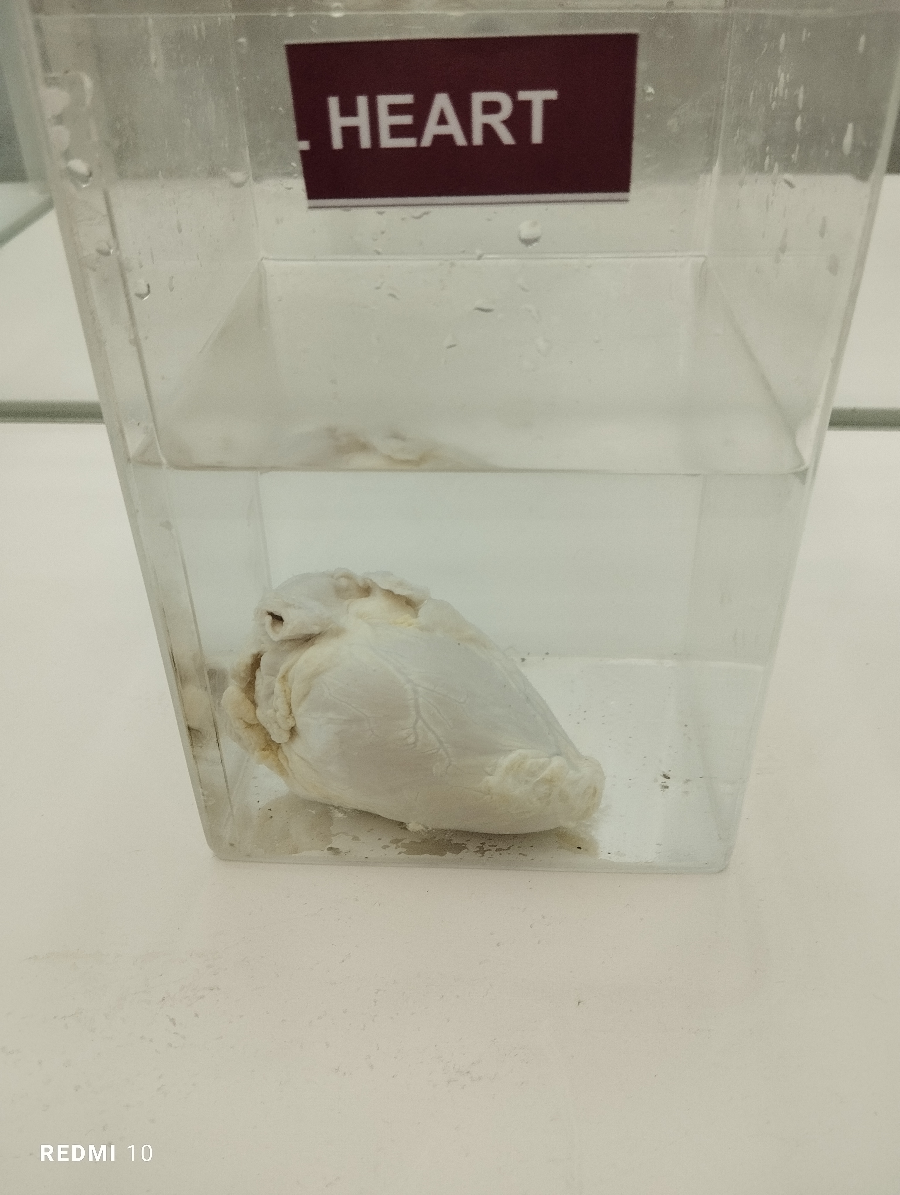
Uterine fibroids, also called leiomyomas or uterine myomas, are benign myometrial tumors of muscle and connective tissue. The incidence of malignancy is 0.36 per 100,000 person-yr. 1 They are typically discrete nodular tumors that vary in size and number.
Fibroids are classified by location in the uterus relative to the myometrium.
•
Subserosal: Located underneath the uterine serosa
•
Intramural: Located in the myometrium proper
•
Submucosal: Also known as intracavitary as they are located adjacent to the endometrium and protrude into the uterine cavity
•
Pedunculated: Located on a pedicle or stalk, either from the serosa or in the cavity
•
The above classifications are further detailed by the FIGO classification system (types 0 to 8)
Fibroids can occur singly but are often multiple. They can be located infrequently within the cervix, broad ligament, adnexa, vagina, vulva, or other unexpected structures. Parasitic fibroids attach to nearby pelvic structures and acquire a blood supply from a nonuterine source with potential detachment from the parent myometrium (Fig. E1). After uncontained morcellation, disseminated peritoneal leiomyomatosis can occur. 1
FIG. E1
Sites of fibroids throughout the uterus.
(From Magowan BA: Clinical obstetrics & gynecology, ed 4, London, 2019, Elsevier.)
SYNONYMS
- Uterine leiomyomas
- Uterine myomas
- Fibroids
View full size
ICD-10CM CODES
D25.0
Submucous leiomyoma of uterus
D25.1
Intramural leiomyoma of uterus
D25.2
Subserosal leiomyoma of uterus
D25.9
Leiomyoma of uterus, unspecified
EPIDEMIOLOGY & DEMOGRAPHICS
•
Estimated cumulative incidence of >70% in women by menopause, generally age 50.
•
50% of fibroids are asymptomatic, but symptomatic uterine fibroids affect 25% to 50% of all women and 30% to 40% of perimenopausal women.
•
Most common benign solid pelvic tumor diagnosed in women and most common reason for benign hysterectomy.
•
Risk factors include age, family history, African American race, early menarche, exposure to diethylstilbestrol, obesity, hypertension, polycystic ovary syndrome, vitamin D deficiency, and nulliparity.
•
Black women typically develop fibroids and clinically significant symptoms at a younger age. 2 The prevalence of fibroids in Black women is 2 to 3 times higher than their White counterparts.
•
Potential to enlarge during pregnancy as well as regress after menopause.
•
Approximately 200,000 hysterectomies, 30,000 myomectomies, thousands of selective uterine-artery embolization and high-intensity focused MRI/ultrasound procedures, and a growing number of radiofrequency ablations are performed annually in the U.S. to remove or destroy uterine fibroids.
PHYSICAL FINDINGS & CLINICAL PRESENTATION
•
Presenting symptoms:
1.
Abnormal uterine bleeding (most common, AUB-L) due to dilated endometrial vessels
2.
Chronic pelvic pain (dysmenorrhea, dyspareunia, pelvic pressure)
3.
Bulk symptoms (bloating, increase in abdominal girth)
4.
Anemia from menorrhagia
5.
Acute pain (torsion of pedunculated fibroid, fibroid infarction or degeneration)
6.
Urinary symptoms (frequency from bladder pressure, ureteral obstruction, urinary retention, incontinence in setting of prolapse)
7.
GI symptoms (rectosigmoid compression with constipation or intestinal obstruction, pain with defecation)
8.
Prolapse through cervix of pedunculated submucosal fibroid
9.
Infertility (sole etiology in 1%-3%, more likely subfertility with submucosal fibroid) 3
10.
Pregnancy complications including preterm birth, small fetus for gestational age, malpresentation
11.
Enlarged, irregular uterus on pelvic examination
ETIOLOGY
Fibroids arise from a single progenitor smooth muscle cell in the myometrium, with recent implication of MED12 somatic mutation as a commonly found potential etiology. They are sensitive to endogenous but not exogenous estrogen and progesterone hormones, thus develop during the reproductive years. Once the fibroids are formed, they promote their own growth via the creation of a local high estrogenic environment. Malignant transformation of preexisting leiomyoma is extremely uncommon (<0.5%). A small group of individuals have an autosomal dominant disorder, hereditary leiomyomatosis and renal cell carcinoma syndrome (HLRCC), in which there is a genetic mutation in the fumarate hydratase gene, causing diminished suppressor function in fibroid formation. 3
DIAGNOSIS
DIFFERENTIAL DIAGNOSIS
•
Ovarian mass (neoplastic, nonneoplastic, endometrioma)
•
Adenomyosis/adenomyoma
•
Endometriosis
•
Endometrial polyp
•
Endometrial carcinoma or hyperplasia
•
Leiomyosarcoma/uterine carcinosarcoma
•
Inflammatory mass (reproductive organ or GI origin)
•
Pregnancy
WORKUP
•
Complete pelvic examination, including speculum exam and bimanual exam
•
Estimation of size of uterus/mass and location of fibroids via imaging
•
Endometrial sampling may be indicated (biopsy or dilation and curettage) when abnormal bleeding and pelvic mass are present to evaluate for endometrial carcinoma; endometrial sampling may identify but cannot rule out leiomyosarcoma, which has no reliable nonsurgical test available
•
Fibroid biopsy not recommended 1
•
If significant urinary symptoms are prominent, intravenous pyelogram to rule out impingement on urinary system
LABORATORY TESTS
•
Pregnancy test
•
CBC
•
Blood urea nitrogen/creatinine
•
Thyroid-stimulating hormone
IMAGING STUDIES
•
Pelvic ultrasound ( Fig. E2 ) is the primary diagnostic modality, with higher diagnostic accuracy via transvaginal ultrasound.
FIG. E2
Fibroid uterus: Endovaginal ultrasound.
Ultrasound is the primary modality used for evaluation of uterine fibroids (leiomyomas). Typical features include a well-circumscribed appearance. Fibroids may be hypoechoic or hyperechoic relative to the uterus. They may be exophytic or intramural, or they may project into the uterine cavity. Whereas malignant uterine tumors may invade adjacent structures, a fibroid is contained within the uterine serosa. Uterine tumors, both benign and malignant, can show central necrosis, which usually appears hypoechoic with ultrasound. In this 38-yr-old woman, the fibroid is exophytic. The right ovary lies adjacent and is difficult to distinguish in this case.
(From Broder JS: Diagnostic imaging for the emergency physician, Philadelphia, 2011, Saunders.)
•
MRI with contrast ( Fig. E3 ) is helpful in planning treatment or if malignancy is strongly suspected. Also relevant to localize fibroid size, number, and location, especially if myomectomy or uterine artery embolism is contemplated.
FIG. E3
Variable appearance of fibroids on magnetic resonance image (MRI).
A, Sagittal T2-weighted MRI showing an intermediate signal anterior myometrial fibroid. Patchy high-signal areas indicate degeneration. A second fibroid at the fundus is of characteristic low signal. B, There is cystic degeneration in the anterior fibroid, and a low signal intensity posterior fibroid that has displaced the rectum. Note retroverted uterus. C, A large pedunculated fibroid is of mixed signal, indicating degeneration. Note multiple small low signal fibroids in the myometrium.
(From Adam A et al: In Grant LA: Grainger & Allison’s diagnostic radiology essentials, ed 2, Philadelphia, 2019, Elsevier.)
•
Saline infusion sonography can be helpful in determining location and protrusion into uterine cavity of submucosal fibroids.
•
Diagnostic hysteroscopy can be performed in the office and may provide direct evidence of intrauterine pathology or submucosal fibroid that distorts uterine cavity.
TREATMENT
Management ( Fig. E4 ) should be based on primary symptoms and patient goals; this may include observation with close follow-up, medical management, temporizing surgical therapies, embolization ( Fig. E5 ), or definitive surgical procedures. Treatment is indicated if bleeding requires blood transfusions, renal function is affected by size of the enlarged fibroid uterus, or when symptoms are present and are severe enough to be unacceptable to the patient.
FIG. E4
Algorithm for the management of uterine fibroids.
AUB, Abnormal uterine bleeding; BSO, bilateral salpingo-oophorectomy; GnRH, gonadotropin-releasing hormone; LNG-IUS, levonorgestrel-releasing intrauterine system; MRg-FUS, magnetic resonance-guided focused ultrasound; OC, oral contraceptives; SPRM, selective progesterone-receptor modulator; UAE, uterine artery embolization.
(From Vilos GA et al: The management of uterine leiomyomas, J Obstet Gynaecol Can 37[2]:163, 2015; and Carranza-Mamane B et al: Society of Obstetrics and Gynaecology Canada Reproductive Endocrinology and Infertility Committee: the management of uterine fibroids in women with otherwise unexplained infertility, SOGC Clinical Practice Guidelines, J Obstet Gynaecol Can 37[3]:277-285, 2015.)
FIG. E5
Uterine fibroid embolization.
A, Angiographic image of uterine leiomyoma before uterine fibroid embolization. Arrows point to preembolization uterine artery. B, Postembolization image of same devascularized myoma with normal myometrial perfusion maintained (black arrows) . White arrow points to patent cervicovaginal branch of uterine artery at completion of embolization.
(From Spies JB, Czeyda-Pommersheim F: Uterine fibroid embolization. In Mauro MA et al [eds]: Image-guided interventions, ed 2, Philadelphia, 2014, Elsevier, pp 542-546. In Gershenson DM et al: Comprehensive gynecology, ed 8, Philadelphia, 2022, Elsevier.)
NONSURGICAL Rx
•
Patient observation and follow-up with periodic repeat pelvic exams to ensure that tumors are not growing rapidly, which could suggest malignancy.
•
Hormonal therapies reduce bleeding symptoms and many have the added benefit of contraception. There is no evidence that exogenous estrogen or progestin increases risk of myomas.
1.
Combined hormonal methods with estrogen and progestin, including oral contraceptives, contraceptive patch, contraceptive vaginal ring
2.
Progestin-only agents including oral progestins, intramuscular progestin injection (DMPA), levonorgestrel intrauterine device (IUD)
a.
Progesterone IUD significantly improves menorrhagia, but expulsion rate with myomas is 10% to 15%, and intracavitary fibroids are a contraindication. Enormous uteri may lead to migrated strings precluding simple retrieval.
b.
A 5-yr DMPA study published in 2022 found reduced fibroid incidence/growth and potential for shrinking fibroid size with use. 4
•
Gonadotropin-releasing hormone (GnRH) agonist, leuprolide, results in 35% to 50% reduction in uterine volume, and 90% to 95% of patients experienced enhanced bleeding control within 3 mo of initiating treatment. Hypoestrogenism, reversible bone loss, and hot flushes are side effects. Consider low-dose progesterone replacement (add-back therapy) to minimize hypoestrogenic effects. It is advised to start add-back therapy within 1 to 3 mo of treatment initiation. GnRH agonist therapy is not recommended for longer than 6 mo without add-back or 12 mo with add-back due to these effects, so goal is to bridge to other treatment. Regrowth and return of bleeding symptoms occur in 50% of treated patients within 3 mo after cessation.
•
Indications for GnRH agonist:
1.
Anemia treatment to normalize hemoglobin before surgery
2.
Avoiding surgery in patients approaching menopause
3.
Preoperatively for large myomas to make hysterectomy or hysteroscopic resection/ablation more feasible
4.
Medical contraindication for surgery
•
Use of GnRH agonists alters the consistency of the fibroid, making myomectomy more challenging.
•
GnRH antagonists (FDA approved 2020 and 2021)—elagolix and relugolix. Both treatment options also have formulations that are combined with hormonal add-back (estradiol and norethindrone acetate) and can be prescribed for max of 2 yr when add-back therapy included (1yr if not). May still have light menses. Possible adverse effects include high blood pressure, worsening of lipids and liver enzymes, and loss of bone mineral density.
•
Nonhormonal medical therapies:
1.
NSAIDs for pain; data does not support use as monotherapy for AUB-L menorrhagia. 3
2.
Tranexamic acid, an oral antifibrinolytic, can decrease menorrhagia by 40% to 65%. Side effects include abdominal cramps, headaches, fatigue, and increased risk of venous thromboembolism.
•
Other drugs used and under investigation:
1.
Mifepristone: Antiprogesterone reduces fibroid volume 40% to 50% with amenorrhea. Benign endometrial changes can occur thus long term data needed
2.
Raloxifene: Selective estrogen receptor modulator, either alone or with GnRH agonist, reduces fibroid volume 70% up to 1 yr but only in postmenopausal patients
SURGICAL Rx
Indications for surgery:
•
Abnormal uterine bleeding with anemia refractory to hormonal therapy
•
Chronic pain with severe dysmenorrhea, dyspareunia, or lower abdominal pressure/pain/compression symptoms
•
Acute pain, torsion, or prolapsing submucosal fibroid
•
Urinary symptoms or signs such as hydronephrosis
•
Infertility or recurrent pregnancy loss with endometrial cavity-distorting fibroid as only finding 5
•
Rapid uterine enlargement premenopausal or any growth after menopause—this indication should involve gynecologic oncologist
•
Procedures:
1.
Hysterectomy (definitive procedure): Vaginal, laparoscopic, robotic, or abdominal approach, dependent on surgical preference/expertise and uterine size.
2.
Myomectomy (preserves fertility): May be performed via abdominal, laparoscopic, or robotic approach; 60% recurrence at 5 yr postop; 6 significant hemorrhage risk, so control anemia preop.
3.
Vaginal myomectomy for prolapsed pedunculated submucosal fibroid.
4.
Hysteroscopic resection: Typically, at least 50% of the fibroid must be intracavitary for a hysteroscopic approach to be successful. May need to be done in stages, as roughly 15% to 20% of patients may need repeat surgery. 6
5.
Endometrial ablation: Decreases menorrhagia, has limitations based on uterine cavity distortion.
6.
Radiofrequency volumetric thermal ablation: FDA-approved uterine-sparing procedure in which laparoscopic or transcervical ultrasound-guided ablation of fibroids induces myolysis (necrosis) resulting in fibroid shrinking. Preliminary data on subsequent pregnancy is limited, but encouraging. 3 , 7
7.
Magnetic resonance–guided focused ultrasound: Ultrasound energy focused to cause fibroid coagulative necrosis. Enables fertility preservation, but due to numerous contraindications, many patients are ineligible. 3
8.
Uterine artery embolization (UAE): Alternative to surgery performed by interventional radiologist, but its less invasive nature should be balanced against a higher rate for treatment failure or complications at 5 yr. Age ≤40 at time of embolization and history of previous myomectomy are predictors of UAE failure. If the patient wishes to preserve future fertility, UAE should not be performed. Contraindicated if history of GnRH agonist use or salpingectomy. 3
•
Patients who receive procedures other than definitive management are more likely to result in a second surgery if younger age/less proximity to menopause
COMPLICATIONS
•
Degeneration occurs when the fibroid outgrows its blood supply. During pregnancy, rare but typically seen in the second trimester. May see fever and leukocytosis, but symptoms improve within 48 h. 3
•
Leiomyosarcoma (<0.1%): The U.S. FDA voiced concern regarding the use of power morcellation in the peritoneal cavity during minimally invasive laparoscopy if a tissue containment system is not used due to the possibility of spreading occult malignancy. In 2020, FDA recommended against power morcellation if age >50 yr or postmenopausal. 1
DISPOSITION
•
Fibroids continue to grow during reproductive years, but symptoms improve after menopause.
•
Early initiation of contraceptive steroids or oral GnRH antagonist combinations in affected person with symptomatic fibroids may reduce the risk of surgery or the extent of surgery.