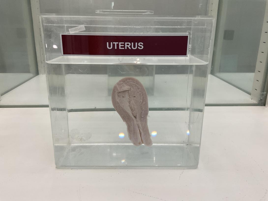
The uterus is a hollow, pear-shaped organ that is an essential part of the female reproductive system. Here are some gross anatomical points related to the uterus:
- Location: The uterus is located in the pelvic cavity, between the bladder and rectum, and is held in place by several ligaments.
- Size and shape: The size and shape of the uterus can vary depending on age and reproductive status. In a non-pregnant adult, the uterus is approximately 7.5 cm long, 5 cm wide, and 2.5 cm thick. It is typically pear-shaped with a broad upper part, the fundus, and a narrow lower part, the cervix.
- Layers: The uterus has three main layers:
- Perimetrium: The outermost layer of the uterus that is a thin layer of connective tissue.
- Myometrium: The middle layer of the uterus that is composed of smooth muscle tissue. The myometrium is responsible for the contractions of the uterus during labor and menstruation.
- Endometrium: The innermost layer of the uterus that is a glandular lining. The endometrium thickens and sheds during the menstrual cycle.
- Structures: The uterus is composed of several structures, including:
- Fundus: The rounded upper part of the uterus that lies above the entrance of the fallopian tubes.
- Body: The main part of the uterus between the fundus and the cervix.
- Cervix: The lower, narrow part of the uterus that connects to the vagina.
- Uterine cavity: The hollow space inside the uterus where the fetus grows during pregnancy.
- Fallopian tubes: Two tubes that extend from the uterus towards the ovaries. The fallopian tubes are the site of fertilization of the egg by the sperm.
Uterine fibroids are noncancerous growths of the uterus that often appear during childbearing years. Also called leiomyomas (lie-o-my-O-muhs) or myomas, uterine fibroids aren't associated with an increased risk of uterine cancer and almost never develop into cancer.
Fibroids range in size from seedlings, undetectable by the human eye, to bulky masses that can distort and enlarge the uterus. You can have a single fibroid or multiple ones. In extreme cases, multiple fibroids can expand the uterus so much that it reaches the rib cage and can add weight.
Many women have uterine fibroids sometime during their lives. But you might not know you have uterine fibroids because they often cause no symptoms. Your doctor may discover fibroids incidentally during a pelvic exam or prenatal ultrasound.
Products & Services
Show more products from Mayo Clinic
Symptoms
Many women who have fibroids don't have any symptoms. In those that do, symptoms can be influenced by the location, size and number of fibroids.
In women who have symptoms, the most common signs and symptoms of uterine fibroids include:
- Heavy menstrual bleeding
- Menstrual periods lasting more than a week
- Pelvic pressure or pain
- Frequent urination
- Difficulty emptying the bladder
- Constipation
- Backache or leg pains
Rarely, a fibroid can cause acute pain when it outgrows its blood supply, and begins to die.
Fibroids are generally classified by their location. Intramural fibroids grow within the muscular uterine wall. Submucosal fibroids bulge into the uterine cavity. Subserosal fibroids project to the outside of the uterus.
When to see a doctor
See your doctor if you have:
- Pelvic pain that doesn't go away
- Overly heavy, prolonged or painful periods
- Spotting or bleeding between periods
- Difficulty emptying your bladder
- Unexplained low red blood cell count (anemia)
Seek prompt medical care if you have severe vaginal bleeding or sharp pelvic pain that comes on suddenly.

