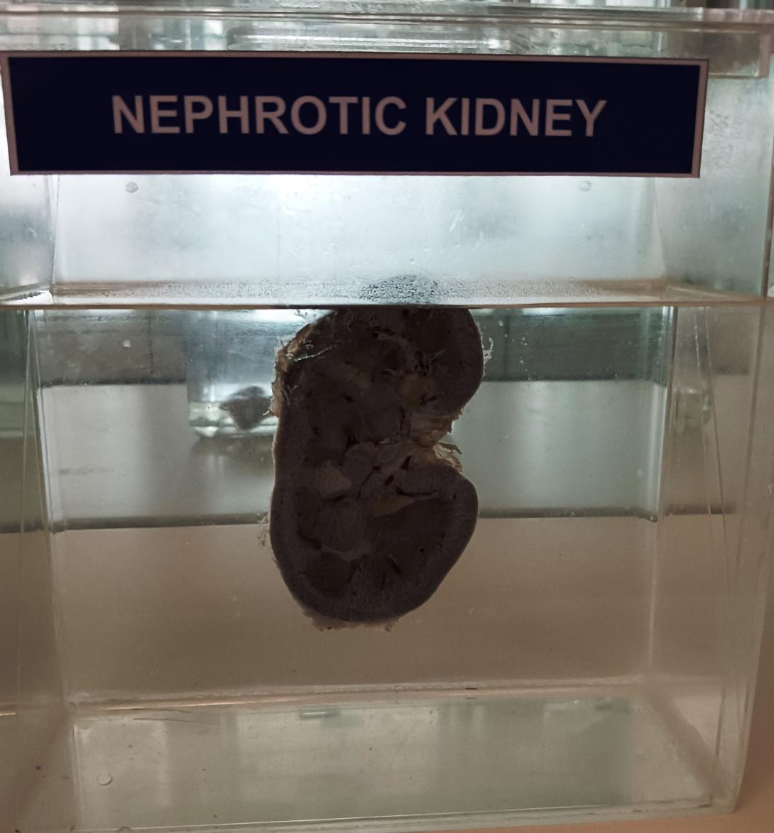
Nephrotic syndrome (NS) is a clinical syndrome defined by massive proteinuria (greater than 40 mg/m^2 per hour) responsible for hypoalbuminemia (less than 30 g/L), with resulting hyperlipidemia, edema, and various complications. It is caused by increased permeability through the damaged basement membrane in the renal glomerulus. It results from an abnormality of glomerular permeability that may be primary with a disease-specific to the kidneys or secondary to congenital infections, diabetes, systemic lupus erythematosus, neoplasia, or certain drug use. There are different types of glomerulonephritis causing nephrotic syndrome, and they all behave differently when it comes to histopathological features of the kidney biopsy.
Minimal change disease is the most common pathology found in childhood (77% to 85%). Usually idiopathic. Light microscopy of renal biopsy samples shows no change; on electron microscopy, effacement of the foot processes can be seen.[31] Immunofluorescent staining for immune complexes is negative.
Focal segmental glomerulosclerosis accounts for 10% to 15% of cases. Light microscopy of renal biopsy sample shows scarring, or sclerosis, of portions of selected glomeruli which can progress into global glomerular sclerosis and tubular atrophy. In most cases, negative immunofluorescence.
Membranoproliferative glomerulonephritis: More commonly presents as nephrotic syndrome. It involves immune complex deposition. Immunofluorescence staining shows a granular pattern. On light microscopy, one can see thickened basement membrane.[32]
Membranous glomerulonephritis: Just 2% to 4% of cases in children, but the most common type in adults. Thickened basement membrane and granular pattern on immunofluorescence. A characteristic “spike and dome” appearance is visible on electron microscopy, with membrane deposition growing around subepithelial immune complex deposition.

