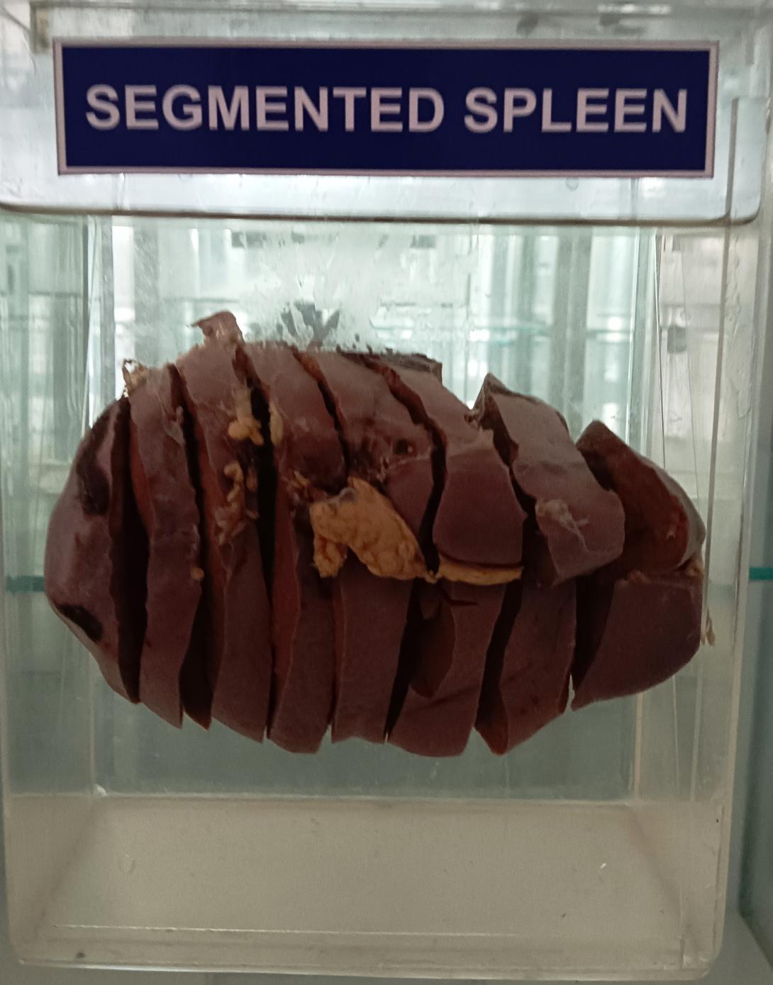
Kyb-r (1870) described the spleen in man, cat, dog, horse and rabbit as being divided into several segments by 'fibrous septa'. He stated that each segment was supplied by its own main artery. Tait & Cashin (1925) confirmed the presence of segments in the spleen of the dog and cat and showed that stimulation of individual neurovascular bundles in the dog spleen produced localized contraction of a segment. However, Dreyer & Budtz-Olson (1952) are commonly regarded as being the first workers to describe the human spleen as consisting of a series of segments each with its own vein: they discovered this while carrying out diagnostic splenic venography. Braithwaite & Adams (1956) studied the splenic segments by injecting radioopaque media and stated that each segment had an independent arterial supply and venous drainage. Clausen (1958) and Gutierrez (1969) demonstrated splenic segments in corrosion casts of the splenic artery and its branches. Likewise, the present paper reports on segmentation in the human spleen after making corrosion casts of the splenic artery and its branches.
Corrosion casts of human splenic arterial trees revealed the presence of two segments-a superior, and an inferior - in 84% of cases and three segments - a superior, a middle and an inferior - in 16% of cases. These segments are separated by avascular planes.
The spleen is an organ found in almost all vertebrates. Similar in structure to a large lymph node, it acts primarily as a blood filter. The word spleen comes from Ancient Greek σπλήν (splḗn).[1]
The spleen plays very important roles in regard to red blood cells (erythrocytes) and the immune system.[2] It removes old red blood cells and holds a reserve of blood, which can be valuable in case of hemorrhagic shock, and also recycles iron. As a part of the mononuclear phagocyte system, it metabolizes hemoglobin removed from senescent red blood cells. The globin portion of hemoglobin is degraded to its constitutive amino acids, and the heme portion is metabolized to bilirubin, which is removed in the liver.[3][4]
The spleen houses antibody-producing lymphocytes in its white pulp and monocytes which remove antibody-coated bacteria and antibody-coated blood cells by way of blood and lymph node circulation. These monocytes, upon moving to injured tissue (such as the heart after myocardial infarction), turn into dendritic cells and macrophages while promoting tissue healing.[5][6][7] The spleen is a center of activity of the mononuclear phagocyte system and is analogous to a large lymph node, as its absence causes a predisposition to certain infections.[8][4]
In humans, the spleen is purple in color and is in the left upper quadrant of the abdomen.[3][9]
Blood supply[edit]
Visceral surface of the spleen
Near the middle of the spleen is a long fissure, the hilum, which is the point of attachment for the gastrosplenic ligament and the point of insertion for the splenic artery and splenic vein. There are other openings present for lymphatic vessels and nerves.
Like the thymus, the spleen possesses only efferent lymphatic vessels. The spleen is part of the lymphatic system. Both the short gastric arteries and the splenic artery supply it with blood.[14]
The germinal centers are supplied by arterioles called penicilliary radicles.[15]

